Martinet Wim, Roth Lynn, De Meyer Guido R Y
Laboratory of Physiopharmacology, University of Antwerp, Universiteitsplein 1, B-2610 Antwerp, Belgium.
Cells. 2017 Jun 30;6(3):17. doi: 10.3390/cells6030017.
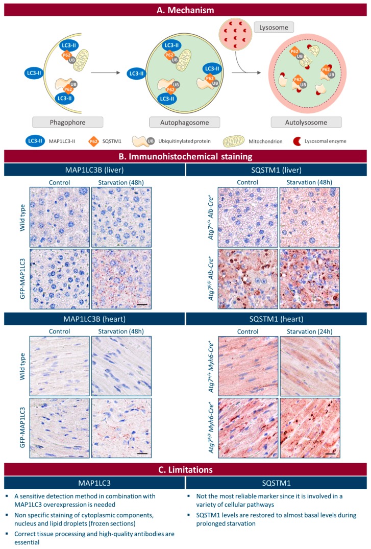
Autophagy is a highly conserved lysosomal degradation pathway with major impact on diverse human pathologies. Despite the development of different methodologies to detect autophagy both in vitro and in vivo, monitoring autophagy in tissue via immunohistochemical techniques is hampered due to the lack of biomarkers. Immunohistochemical detection of a punctate pattern of ATG8/MAP1LC3 proteins is currently the most frequently used approach to detect autophagy in situ, but it depends on a highly sensitive detection method and is prone to misinterpretation. Moreover, reliable MAP1LC3 immunohistochemical staining requires correct tissue processing and high-quality, isoform-specific antibodies. Immunohistochemical analysis of other autophagy-related protein targets such as SQSTM1, ubiquitin, ATG5 or lysosomal proteins is not recommended as marker for autophagic activity in tissue for multiple reasons including aspecific labeling of cellular structures and a lack of differential protein expression during autophagy initiation. To better understand the role of autophagy in human disease, novel biomarkers for visualization of the autophagic process with standard histology techniques are urgently needed.
自噬是一种高度保守的溶酶体降解途径,对多种人类疾病具有重大影响。尽管已经开发出不同的方法在体外和体内检测自噬,但由于缺乏生物标志物,通过免疫组织化学技术在组织中监测自噬受到阻碍。目前,免疫组织化学检测ATG8/MAP1LC3蛋白的点状模式是在原位检测自噬最常用的方法,但它依赖于高度灵敏的检测方法,并且容易产生误解。此外,可靠的MAP1LC3免疫组织化学染色需要正确的组织处理和高质量的、亚型特异性抗体。不建议将其他自噬相关蛋白靶点(如SQSTM1、泛素、ATG5或溶酶体蛋白)的免疫组织化学分析作为组织中自噬活性的标志物,原因有多种,包括细胞结构的非特异性标记以及自噬起始过程中缺乏差异蛋白表达。为了更好地理解自噬在人类疾病中的作用,迫切需要能够用标准组织学技术可视化自噬过程的新型生物标志物。