Ohmiya Naoki, Horiguchi Noriyuki, Tahara Tomomitsu, Nagasaka Mitsuo, Nakagawa Yoshihito, Shibata Tomoyuki, Tsukamoto Tetsuya, Kuroda Makoto
Department of Gastroenterology, Fujita Health University School of Medicine, Toyoake, Aichi, Japan.
Department of Diagnostic Pathology I, Fujita Health University School of Medicine, Toyoake, Aichi, Japan.
Endosc Int Open. 2017 Jul;5(7):E547-E558. doi: 10.1055/s-0043-106184. Epub 2017 Jun 23.
Probe-based confocal laser endomicroscopy (pCLE) enables real-time optical biopsy. Little is known about pCLE imaging deep inside the small bowel, therefore the aim of this study was to determine its usefulness.
Between April 2014 and January 2016, we performed 38 pCLE examinations during double-balloon enteroscopy with intravenous fluorescein in 37 patients with: tumors (n = 10), vascular disorders (n = 6), inflammatory diseases and drug injuries (n = 13), other disorders (n = 4), and normal findings (n = 4). We measured the calibers of capillary and lymphatic vessels at 15 different sites and compared the calibers between pCLE images and histopathology. We also compared pCLE findings with pathologic diagnosis.
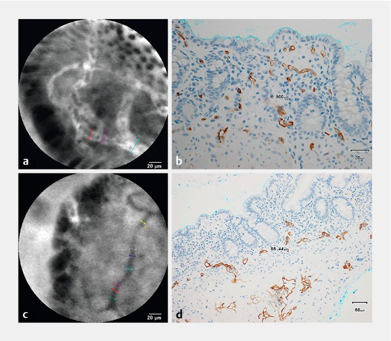
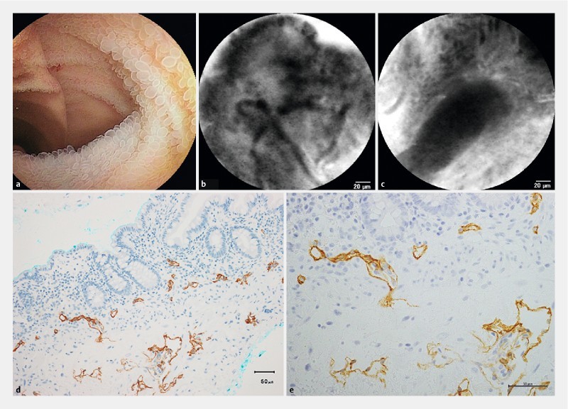
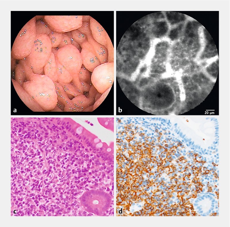
The inner diameters of capillary vessels beneath the epithelium and in the middle of villi were 16.2 ± 3.0 µm and 14.5 ± 3.1 µm, respectively, in the pCLE images, but these were not consistent with formalin-fixed paraffin-embedded histology. In tumors, larger capillary vessels were observed in aggressive malignant lymphoma and metastasis, and a "soccer ball-like pattern" constituting homogenous dark cells packed with polygonal, narrower capillary vessels was characteristic in pCLE images of a malignant lymphoma follicle. Hereditary hemorrhagic telangiectasia and angiodysplasia contained anastomosis of capillary vessels of different calibers. In IgA vasculitis, segmental capillary strictures were observed. Intestinal lymphangiectasia with protein-losing enteropathy contained a reticular network of lymphatic vessels and dilated lymphatic ducts accompanied by numerous cell gaps. pCLE findings corresponded to pathologic diagnosis in 32 examinations (91 %).
pCLE is useful for in vivo analysis of abnormalities of the capillary and lymphatic vessels and epithelium, and for diagnosis in various small-bowel diseases.
基于探头的共聚焦激光内镜检查(pCLE)可实现实时光学活检。对于小肠深部的pCLE成像了解甚少,因此本研究的目的是确定其效用。
2014年4月至2016年1月期间,我们在37例患者的双气囊小肠镜检查过程中,静脉注射荧光素后进行了38次pCLE检查,这些患者包括:肿瘤(n = 10)、血管疾病(n = 6)、炎症性疾病和药物损伤(n = 13)、其他疾病(n = 4)以及检查结果正常(n = 4)。我们在15个不同部位测量了毛细血管和淋巴管的管径,并比较了pCLE图像与组织病理学之间的管径。我们还将pCLE检查结果与病理诊断进行了比较。
在pCLE图像中,上皮下方和绒毛中部的毛细血管内径分别为16.2±3.0μm和14.5±3.1μm,但这些与福尔马林固定石蜡包埋组织学不一致。在肿瘤中,侵袭性恶性淋巴瘤和转移瘤中观察到较大的毛细血管,恶性淋巴瘤滤泡的pCLE图像中,一种由充满多边形、较窄毛细血管的均匀暗细胞组成的“足球样图案”具有特征性。遗传性出血性毛细血管扩张症和血管发育异常包含不同管径毛细血管的吻合。在IgA血管炎中,观察到节段性毛细血管狭窄。伴有蛋白丢失性肠病的肠淋巴管扩张症包含淋巴管的网状网络和扩张的淋巴管,伴有大量细胞间隙。32次检查(91%)的pCLE检查结果与病理诊断相符。
pCLE可用于对毛细血管、淋巴管和上皮异常进行体内分析,以及用于各种小肠疾病的诊断。