Setiawati Rosy, Utomo Dwikora Novembri, Rantam Fedik Abdul, Ifran Nadia Nastassia, Budhiparama Nicolaas C
Musculoskeletal Division, Department of Radiology, School of Medicine, Airlangga University, Dr Soetomo Hospital, Airlangga University Hospital, Surabaya, Indonesia.
Stem Cell Laboratory, Institute of Tropical Disease, Airlangga University, Surabaya, Indonesia.
Orthop J Sports Med. 2017 Jun 21;5(6):2325967117708548. doi: 10.1177/2325967117708548. eCollection 2017 Jun.
Bone marrow mesenchymal stem cells (BM-MSCs) are multipotent adult stem cells and have become an important source of cells for engineering tissue repair and cell therapy. Vascular endothelial growth factor (VEGF) promotes angiogenesis and contributes fibrous integration between tendon and bone during the early postoperative stage. Both MSCs and VEGF can stimulate cell proliferation, differentiation, and matrix deposition by enhancing angiogenesis and osteogenesis of the graft in the tunnel.
Injection of intratunnel BM-MSCs and VEGF enhances the early healing process of a tendon graft.
Controlled laboratory study.
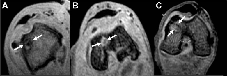
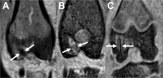
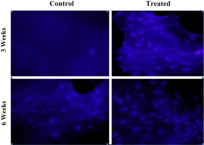
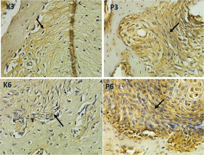
In this controlled animal laboratory study, each of 4 groups of rabbits underwent unilateral anterior cruciate ligament (ACL) reconstruction with use of the ipsilateral semitendinosus tendon. The rabbits received intratunnel injection of BM-MSCs and VEGF with a fibrin glue seal covering the distal tunnel at the articular site. Evaluation using magnetic resonance imaging (MRI), collagen type III expression, and biomechanical analyses were performed at 3- and 6-week intervals.
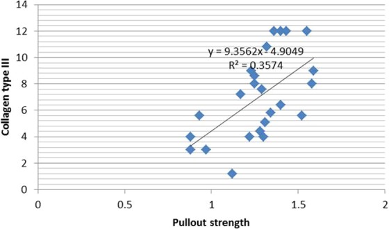
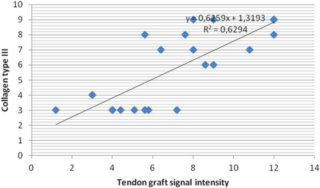
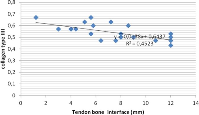
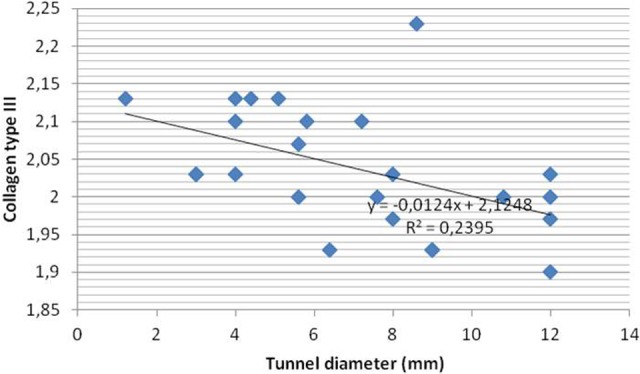
All parameters using MRI, collagen type III expression, and biomechanical analysis of pullout strength of the graft showed that application of intratunnel BM-MSCs and VEGF enhanced tendon-to-bone healing after ACL reconstruction.
Intratunnel injections of BM-MSCs and VEGF after ACL reconstruction enhanced graft tunnel healing. Overall, the femoral tunnel that received BM-MSCs and VEGF had better advanced healing with increased collagen type III fibers and better outcomes on MRI and biomechanical analysis. MRI is the most reliable tool for clinical use in evaluating stages of ACL healing after reconstruction, since biopsy is an invasive procedure.
骨髓间充质干细胞(BM-MSCs)是多能成体干细胞,已成为工程组织修复和细胞治疗的重要细胞来源。血管内皮生长因子(VEGF)可促进血管生成,并在术后早期促进肌腱与骨之间的纤维整合。骨髓间充质干细胞和血管内皮生长因子均可通过增强移植物在隧道内的血管生成和成骨作用来刺激细胞增殖、分化和基质沉积。
隧道内注射骨髓间充质干细胞和血管内皮生长因子可促进肌腱移植物的早期愈合过程。
对照实验室研究。
在这项对照动物实验室研究中,4组兔子均使用同侧半腱肌进行单侧前交叉韧带(ACL)重建。兔子接受隧道内注射骨髓间充质干细胞和血管内皮生长因子,并用纤维蛋白胶密封覆盖关节部位的远端隧道。分别在3周和6周时进行磁共振成像(MRI)评估、III型胶原表达评估和生物力学分析。
使用MRI、III型胶原表达以及移植物拔出强度的生物力学分析的所有参数均显示,隧道内应用骨髓间充质干细胞和血管内皮生长因子可增强ACL重建术后肌腱与骨的愈合。
ACL重建术后隧道内注射骨髓间充质干细胞和血管内皮生长因子可促进移植物隧道愈合。总体而言,接受骨髓间充质干细胞和血管内皮生长因子的股骨隧道愈合进展更好,III型胶原纤维增加,在MRI和生物力学分析方面结果更佳。由于活检是一种侵入性操作,因此MRI是评估ACL重建术后愈合阶段临床应用中最可靠的工具。