Misra Paraish, Kirpalani Anish, Leung General, Vlachou Paraskevi A, Lee Jason Y, Jothy Serge, Zaltzman Jeffrey, Yuen Darren A
Division of Nephrology, St. Michael's Hospital, University of Toronto, Toronto, ON, M5B 1W8, Canada.
Department of Medical Imaging, St. Michael's Hospital, University of Toronto, 30 Bond Street, 3 Cardinal Carter South, Toronto, ON, M5B 1W8, Canada.
BMC Nephrol. 2017 Jul 10;18(1):224. doi: 10.1186/s12882-017-0618-2.
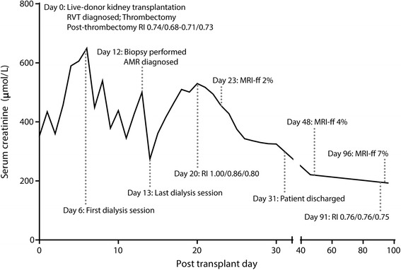
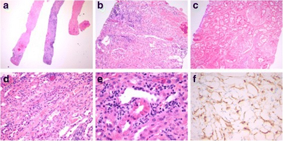
Surgical thrombectomy in the context of acute renal vein thrombosis (RVT) post-transplantation has had limited success, with considerable variation in the surgical techniques used. Unfortunately, it is usually followed by allograft nephrectomy within a few days if rapid allograft recovery does not ensue. We report a case of acute RVT in which nephrectomy was not performed despite a prolonged requirement for dialysis post-thrombectomy, but with recovery of renal function 2 weeks later. We also report the findings of serial MRI with diffusion-weighted imaging (DW-MRI) throughout the patient's recovery, which provided novel insights into allograft microvascular perfusion changes post-thrombectomy.
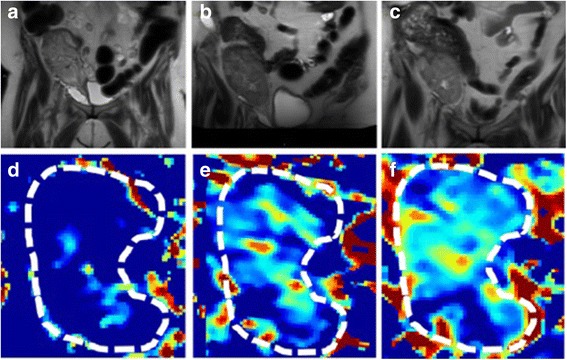
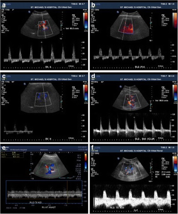
A 65-year old patient underwent living-unrelated kidney transplantation complicated by acute RVT. Surgical thrombectomy and irrigation led to a delayed, but significant, recovery of renal function. Serial non-contrast DW-MRI scanning was used to non-invasively assess microvascular renal blood flow post-operatively. Unlike standard Doppler ultrasonography, DW-MRI documented reduced microvascular perfusion initially, with gradual but incomplete recovery that mirrored the partial improvement in renal function.
Our findings suggest that surgical thrombectomy may be more effective than previously described if followed by careful patient observation. Moreover, diffusion-weighted MRI appears to provide important insights into the pathophysiology of delayed graft function and deserves further investigation.
移植后急性肾静脉血栓形成(RVT)情况下的手术取栓成功率有限,所使用的手术技术差异很大。不幸的是,如果移植肾不能迅速恢复,通常在几天内就会进行移植肾切除术。我们报告一例急性RVT病例,尽管取栓术后需要长期透析,但未进行肾切除术,而是在2周后肾功能恢复。我们还报告了患者整个恢复过程中连续进行的磁共振成像(MRI)及弥散加权成像(DW-MRI)的结果,这些结果为取栓术后移植肾微血管灌注变化提供了新的见解。
一名65岁患者接受了非亲属活体肾移植,并发急性RVT。手术取栓和冲洗导致肾功能延迟但显著恢复。连续非增强DW-MRI扫描用于术后无创评估肾微血管血流。与标准多普勒超声不同,DW-MRI显示最初微血管灌注减少,随后逐渐但不完全恢复,这与肾功能的部分改善情况相符。
我们的研究结果表明,如果术后对患者进行仔细观察,手术取栓可能比之前描述的更有效。此外,弥散加权MRI似乎为移植肾功能延迟的病理生理学提供了重要见解,值得进一步研究。