Centre National de la Recherche Scientifique (CNRS), Université Paris-Sud, Université Paris-Saclay UMR 9199, Neurodegenerative Diseases Laboratory, F-92260, Fontenay-aux-Roses, France.
Commissariat à l'Energie Atomique et aux Energies Alternatives (CEA), Direction de la Recherche Fondamentale (DRF), Molecular Imaging Research Center (MIRCen), F-92260, Fontenay-aux-Roses, France.
Sci Rep. 2017 Jul 10;7(1):4955. doi: 10.1038/s41598-017-05285-1.
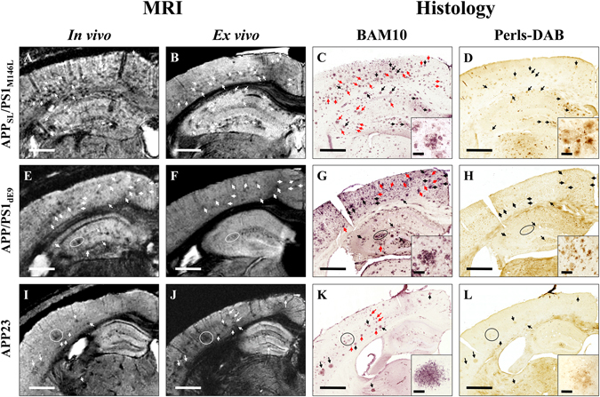
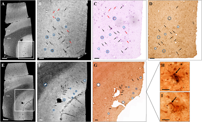
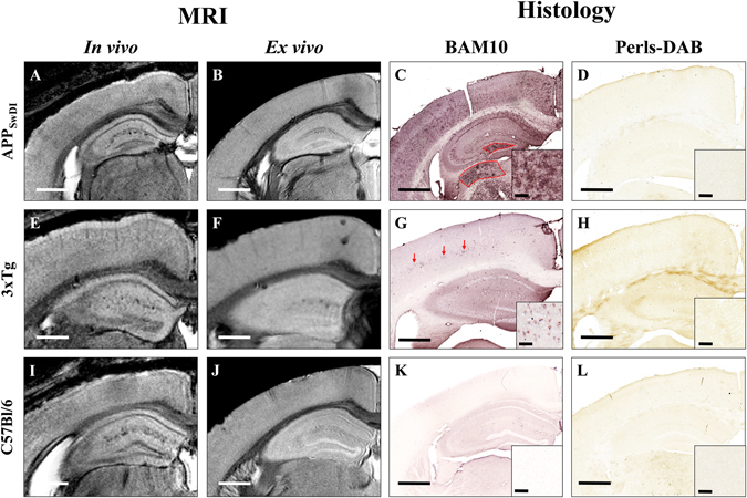
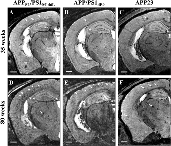
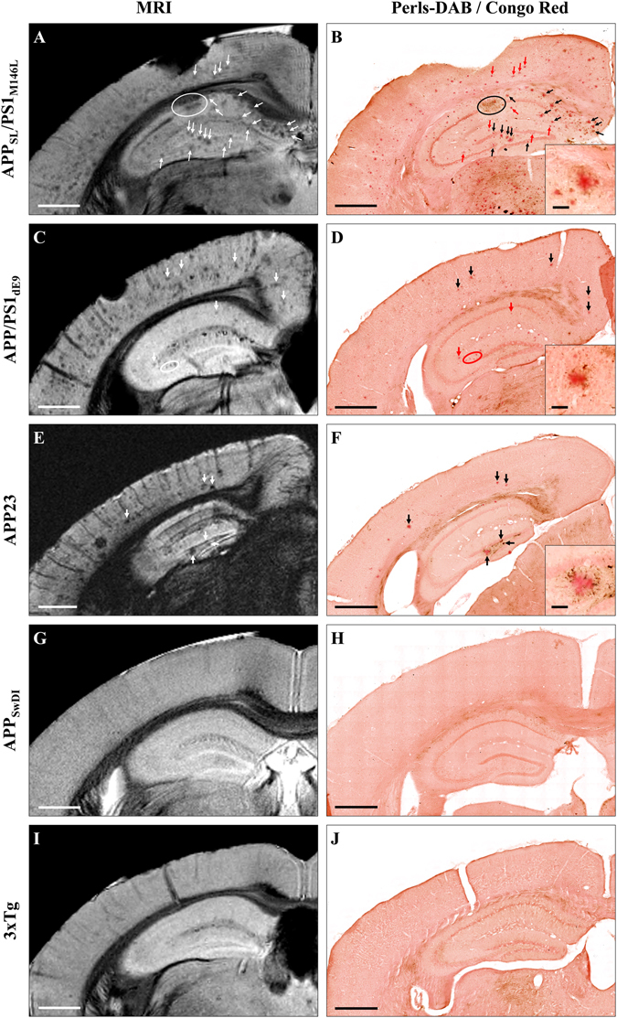
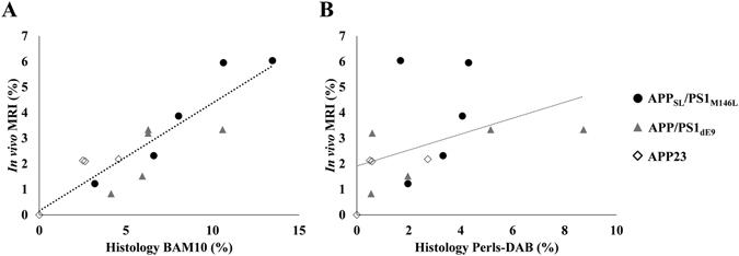
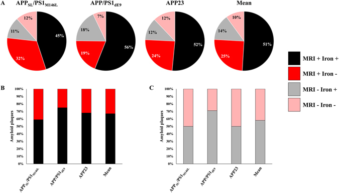
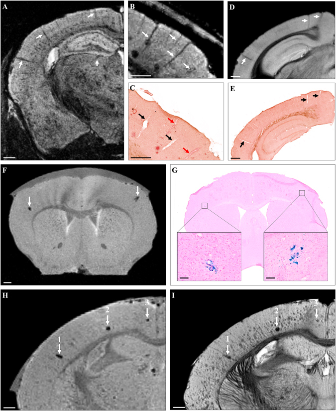
Gadolinium (Gd)-stained MRI is based on Gd contrast agent (CA) administration into the brain parenchyma. The strong signal increase induced by Gd CA can be converted into resolution enhancement to record microscopic MR images. Moreover, inhomogeneous distribution of the Gd CA in the brain improves the contrast between different tissues and provides new contrasts in MR images. Gd-stained MRI detects amyloid plaques, one of the microscopic lesions of Alzheimer's disease (AD), in APP/PS1 mice or in primates. Numerous transgenic mice with various plaque typologies have been developed to mimic cerebral amyloidosis and comparison of plaque detection between animal models and humans with new imaging methods is a recurrent concern. Here, we investigated detection of amyloid plaques by Gd-stained MRI in five mouse models of amyloidosis (APP/PS1, APP/PS1, APP23, APP, and 3xTg) presenting with compact, diffuse and intracellular plaques as well as in post mortem human-AD brains. The brains were then evaluated by histology to investigate the impact of size, compactness, and iron load of amyloid plaques on their detection by MRI. We show that Gd-stained MRI allows detection of compact amyloid plaques as small as 25 µm, independently of their iron load, in mice as well as in human-AD brains.
钆染色磁共振成像(Gd-stained MRI)基于将 Gd 造影剂(CA)注入脑实质。Gd CA 引起的强信号增加可以转化为分辨率增强,以记录微观磁共振图像。此外,Gd CA 在脑内的不均匀分布提高了不同组织之间的对比度,并在磁共振图像中提供了新的对比。Gd 染色 MRI 可检测 APP/PS1 小鼠或灵长类动物阿尔茨海默病(AD)的一种微观病变——淀粉样斑块。已经开发出了许多具有不同斑块类型的转基因小鼠来模拟脑淀粉样变性,并且使用新的成像方法比较动物模型和人类之间的斑块检测情况一直是人们关注的焦点。在这里,我们研究了 Gd 染色 MRI 在五种淀粉样变性小鼠模型(APP/PS1、APP/PS1、APP23、APP 和 3xTg)中检测淀粉样斑块的情况,这些模型具有致密、弥散和细胞内斑块。然后,通过组织学评估这些脑,以研究淀粉样斑块的大小、致密程度和铁负荷对其 MRI 检测的影响。我们表明,Gd 染色 MRI 可检测到直径小至 25 µm 的致密淀粉样斑块,无论其铁负荷如何,在小鼠以及人类 AD 脑中均如此。