Tootell Roger B H, Nasr Shahin
Martinos Center for Biomedical Imaging, Charlestown, Massachusetts 02129, and
Department of Radiology, Harvard Medical School, Boston, Massachusetts 02115.
J Neurosci. 2017 Aug 16;37(33):8014-8032. doi: 10.1523/JNEUROSCI.0690-17.2017. Epub 2017 Jul 19.
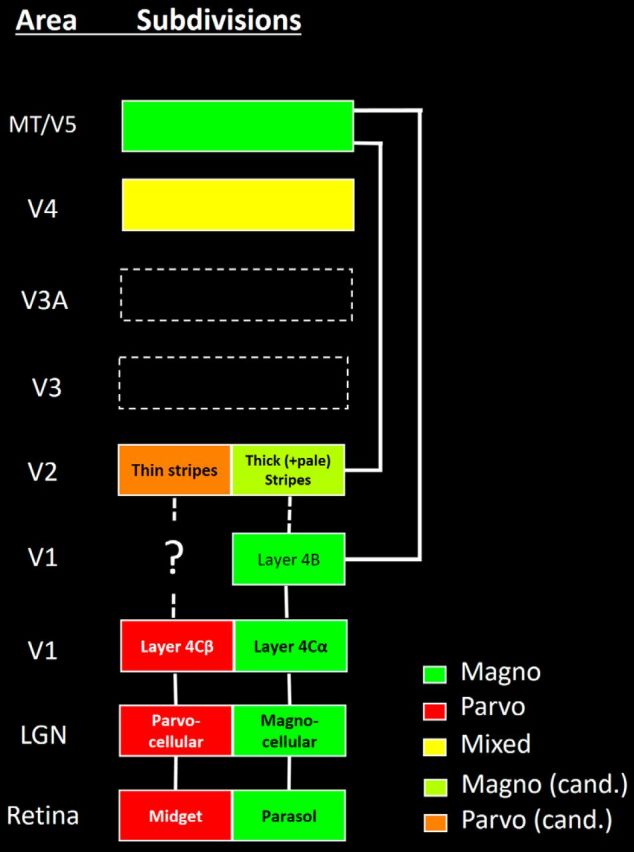
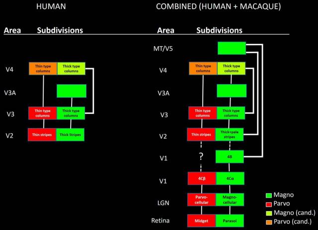
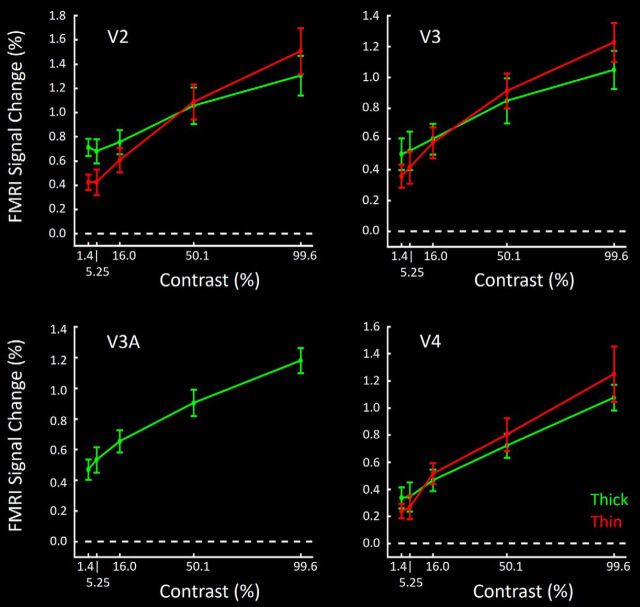
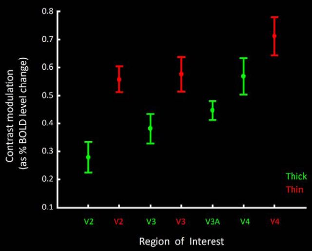
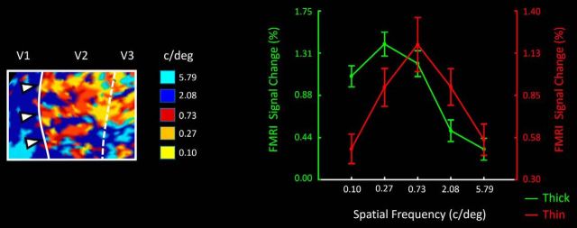
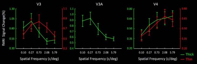
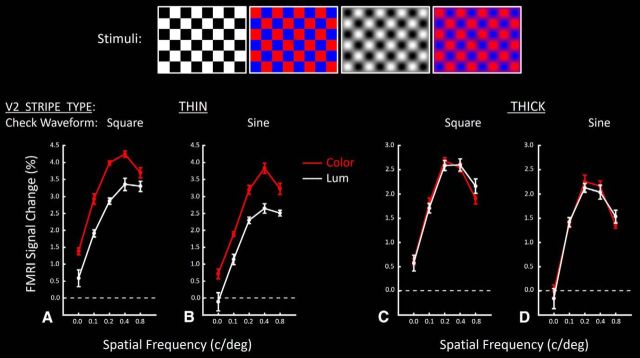
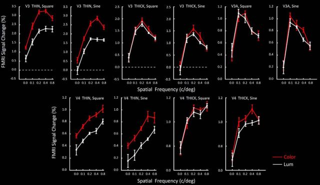
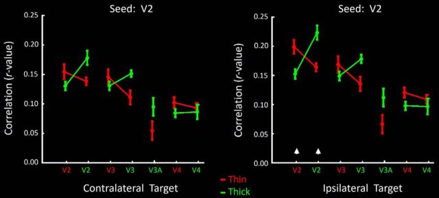
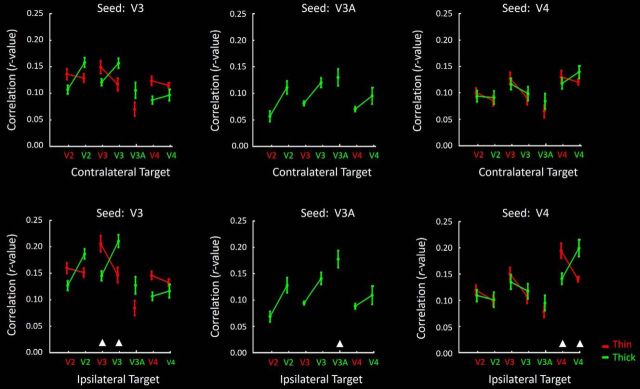
Magnocellular versus parvocellular (M-P) streams are fundamental to the organization of macaque visual cortex. Segregated, paired M-P streams extend from retina through LGN into V1. The M stream extends further into area V5/MT, and parts of V2. However, elsewhere in visual cortex, it remains unclear whether M-P-derived information (1) becomes intermixed or (2) remains segregated in M-P-dominated columns and neurons. Here we tested whether M-P streams exist in extrastriate cortical columns, in 8 human subjects (4 female). We acquired high-resolution fMRI at high field (7T), testing for M- and P-influenced columns within each of four cortical areas (V2, V3, V3A, and V4), based on known functional distinctions in M-P streams in macaque: (1) color versus luminance, (2) binocular disparity, (3) luminance contrast sensitivity, (4) peak spatial frequency, and (5) color/spatial interactions. Additional measurements of resting state activity (eyes closed) tested for segregated functional connections between these columns. We found M- and P-like functions and connections within and between segregated cortical columns in V2, V3, and (in most experiments) area V4. Area V3A was dominated by the M stream, without significant influence from the P stream. These results suggest that M-P streams exist, and extend through, specific columns in early/middle stages of human extrastriate cortex. The magnocellular and parvocellular (M-P) streams are fundamental components of primate visual cortical organization. These streams segregate both anatomical and functional properties in parallel, from retina through primary visual cortex. However, in most higher-order cortical sites, it is unknown whether such M-P streams exist and/or what form those streams would take. Moreover, it is unknown whether M-P streams exist in human cortex. Here, fMRI evidence measured at high field (7T) and high resolution revealed segregated M-P streams in four areas of human extrastriate cortex. These results suggest that M-P information is processed in segregated parallel channels throughout much of human visual cortex; the M-P streams are more than a convenient sorting property in earlier stages of the visual system.
大细胞与小细胞(M-P)信息流对于猕猴视觉皮层的组织架构至关重要。分离的、成对的M-P信息流从视网膜经外侧膝状体核(LGN)延伸至V1。M信息流进一步延伸至V5/MT区以及V2区的部分区域。然而,在视觉皮层的其他区域,源自M-P的信息是(1)相互混合,还是(2)在以M-P为主导的柱体和神经元中保持分离,仍不清楚。在此,我们对8名人类受试者(4名女性)进行测试,以探究M-P信息流是否存在于纹外皮层柱体中。我们在高场强(7T)下采集高分辨率功能磁共振成像(fMRI)数据,基于猕猴M-P信息流中已知的功能差异,在四个皮层区域(V2、V3、V3A和V4)中的每个区域内测试受M和P影响的柱体:(1)颜色与亮度,(2)双眼视差,(3)亮度对比敏感度,(4)峰值空间频率,以及(5)颜色/空间相互作用。对静息状态活动(闭眼)的额外测量测试了这些柱体之间分离的功能连接。我们在V2、V3以及(在大多数实验中)V4区的分离皮层柱体内部及之间发现了类似M和P的功能及连接。V3A区主要由M信息流主导,几乎不受P信息流的影响。这些结果表明,M-P信息流存在,并贯穿人类纹外皮层早期/中期的特定柱体。大细胞和小细胞(M-P)信息流是灵长类动物视觉皮层组织的基本组成部分。这些信息流从视网膜到初级视觉皮层,在解剖学和功能特性上均并行分离。然而,在大多数高阶皮层区域,尚不清楚此类M-P信息流是否存在以及/或者这些信息流会呈现何种形式。此外,人类皮层中是否存在M-P信息流也不清楚。在此,在高场强(7T)和高分辨率下测量的fMRI证据揭示了人类纹外皮层四个区域中分离的M-P信息流。这些结果表明,M-P信息在人类视觉皮层的大部分区域通过分离的并行通道进行处理;M-P信息流不仅仅是视觉系统早期阶段一种方便的分类特性。