• 文献检索
  • 文档翻译
  • 深度研究
  • 学术资讯
  • Suppr Zotero 插件Zotero 插件
  • 邀请有礼
  • 套餐&价格
  • 历史记录
应用&插件
Suppr Zotero 插件Zotero 插件浏览器插件Mac 客户端Windows 客户端微信小程序
定价
高级版会员购买积分包购买API积分包
服务
文献检索文档翻译深度研究API 文档MCP 服务
关于我们
关于 Suppr公司介绍联系我们用户协议隐私条款
关注我们

Suppr 超能文献

核心技术专利:CN118964589B侵权必究
粤ICP备2023148730 号-1Suppr @ 2026

文献检索

告别复杂PubMed语法,用中文像聊天一样搜索,搜遍4000万医学文献。AI智能推荐,让科研检索更轻松。

立即免费搜索

文件翻译

保留排版,准确专业,支持PDF/Word/PPT等文件格式,支持 12+语言互译。

免费翻译文档

深度研究

AI帮你快速写综述,25分钟生成高质量综述,智能提取关键信息,辅助科研写作。

立即免费体验

颞下颌关节锥形束计算机断层扫描图像上疼痛与退行性骨质改变之间的相关性

Correlation between pain and degenerative bony changes on cone-beam computed tomography images of temporomandibular joints.

作者信息

Bae SunMee, Park Moon-Soo, Han Jin-Woo, Kim Young-Jun

机构信息

Department of Oral Medicine and Diagnosis, Research Institute of Oral Science, College of Dentistry, Gangneung-Wonju National University, 7 Jukhyun-gil, Gangneung, 25457 South Korea.

Department of Oral and Maxillofacial Radiology, Research Institute of Oral Science, College of Dentistry, Gangneung-Wonju National University, Gangneung, South Korea.

出版信息

Maxillofac Plast Reconstr Surg. 2017 Jul 5;39(1):19. doi: 10.1186/s40902-017-0117-1. eCollection 2017 Dec.

DOI:10.1186/s40902-017-0117-1
PMID:28730147
原文链接:https://pmc.ncbi.nlm.nih.gov/articles/PMC5496923/
Abstract

BACKGROUND

The aim of this study was to assess correlation between pain and degenerative bony changes on cone-beam computed tomography (CBCT) images of temporomandibular joints (TMJs).

METHODS

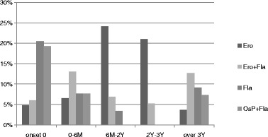
Two hundred eighty-three temporomandibular joints with degenerative bony changes were evaluated. Pain intensity (numeric rating scale, NRS) and pain duration in patients with degenerative joint disease (DJD) were also analyzed. We classified condylar bony changes on CBCT into five types: osteophyte (Osp), erosion (Ero), flattening (Fla), subchondral sclerosis (Scl), and pseudocyst (Pse).

RESULTS

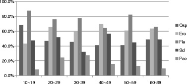
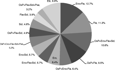
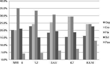
Degenerative bony changes were the most frequent in the age groups of 1019, 20-29, and 5059 years. The most frequent pain intensity was "none" (NRS 0, 34.6%) followed by "annoying" (NRS 3-5, 29.7%). The most frequent condylar bony change was Fla (219 joints, 77.4%) followed by Ero (169 joints, 59.7%). "Ero + Fla" was the most common combination of the bony changes (12.7%). The frequency of erosion was directly proportional to NRS, but the frequency of osteophyte was inversely proportional. The prevalence of Ero increased from onset until 2 years and gradually decreased thereafter. The prevalence of Osp, Ero, and Pse increased with age.

CONCLUSIONS

Osp and Ero can be pain-related variables in degenerative joint disease (DJD) patients. "Six months to 2 years" may be a meaningful time point from the active, unstable phase to the stabilized late phase of DJD.

摘要

背景

本研究旨在评估颞下颌关节(TMJ)锥形束计算机断层扫描(CBCT)图像上疼痛与骨质退行性改变之间的相关性。

方法

对283个存在骨质退行性改变的颞下颌关节进行评估。还分析了退行性关节病(DJD)患者的疼痛强度(数字评定量表,NRS)和疼痛持续时间。我们将CBCT上髁突骨质改变分为五种类型:骨赘(Osp)、侵蚀(Ero)、扁平(Fla)、软骨下硬化(Scl)和假性囊肿(Pse)。

结果

骨质退行性改变在1019岁、2029岁和50~59岁年龄组中最为常见。最常见的疼痛强度为“无”(NRS 0,34.6%),其次是“烦人”(NRS 3 - 5,29.7%)。最常见的髁突骨质改变是Fla(219个关节,77.4%),其次是Ero(169个关节,59.7%)。“Ero + Fla”是最常见的骨质改变组合(12.7%)。侵蚀的频率与NRS成正比,但骨赘的频率成反比。侵蚀的患病率从发病起至2年增加,此后逐渐下降。Osp、Ero和Pse的患病率随患病率随年龄增加。

结论

Osp和Ero可能是退行性关节病(DJD)患者与疼痛相关的变量。“6个月至2年”可能是DJD从活跃、不稳定阶段到稳定后期的一个有意义的时间点。

https://cdn.ncbi.nlm.nih.gov/pmc/blobs/8586/5496923/0d59fe9a4f29/40902_2017_117_Fig5_HTML.jpg
https://cdn.ncbi.nlm.nih.gov/pmc/blobs/8586/5496923/78c8484fd41c/40902_2017_117_Fig1_HTML.jpg
https://cdn.ncbi.nlm.nih.gov/pmc/blobs/8586/5496923/71a9ba12c039/40902_2017_117_Fig2_HTML.jpg
https://cdn.ncbi.nlm.nih.gov/pmc/blobs/8586/5496923/bd2f43a7956a/40902_2017_117_Fig3_HTML.jpg
https://cdn.ncbi.nlm.nih.gov/pmc/blobs/8586/5496923/4ef209c3c96c/40902_2017_117_Fig4_HTML.jpg
https://cdn.ncbi.nlm.nih.gov/pmc/blobs/8586/5496923/0d59fe9a4f29/40902_2017_117_Fig5_HTML.jpg

相似文献

1
Correlation between pain and degenerative bony changes on cone-beam computed tomography images of temporomandibular joints.颞下颌关节锥形束计算机断层扫描图像上疼痛与退行性骨质改变之间的相关性
Maxillofac Plast Reconstr Surg. 2017 Jul 5;39(1):19. doi: 10.1186/s40902-017-0117-1. eCollection 2017 Dec.
2
Condylar bony changes in patients with temporomandibular disorders: a CBCT study.颞下颌关节紊乱病患者髁突骨质改变:一项锥形束计算机断层扫描研究
Imaging Sci Dent. 2012 Dec;42(4):249-53. doi: 10.5624/isd.2012.42.4.249. Epub 2012 Dec 23.
3
Temporomandibular joint osteoarthritis: cone beam computed tomography findings, clinical features, and correlations.颞下颌关节骨关节炎:锥形束计算机断层扫描结果、临床特征及相关性
Int J Oral Maxillofac Surg. 2015 Oct;44(10):1268-74. doi: 10.1016/j.ijom.2015.06.023. Epub 2015 Jul 17.
4
Differences in clinical and radiographic features between bilateral and unilateral adult degenerative temporomandibular joint disease: A retrospective cross-sectional study.双侧和单侧成人退行性颞下颌关节疾病的临床和影像学特征差异:一项回顾性横断面研究。
Int Orthod. 2023 Jun;21(2):100731. doi: 10.1016/j.ortho.2023.100731. Epub 2023 Feb 11.
5
Assessment of bony changes in temporomandibular joint in patients using cone beam computed tomography - a cross sectional study.利用锥形束 CT 评估颞下颌关节的骨改变 - 一项横断面研究。
Head Face Med. 2023 Oct 28;19(1):47. doi: 10.1186/s13005-023-00392-z.
6
Bone changes in the temporomandibular joints of older adults: A cone-beam computed tomography study.老年人颞下颌关节的骨质改变:一项锥形束计算机断层扫描研究。
Spec Care Dentist. 2020 Jan;40(1):84-89. doi: 10.1111/scd.12441. Epub 2019 Dec 4.
7
Bony deviations revealed by cone beam computed tomography of the temporomandibular joint in subjects without ongoing pain.无持续性疼痛患者的颞下颌关节锥形束 CT 检查所显示的骨性偏斜。
J Oral Facial Pain Headache. 2014 Fall;28(4):331-7. doi: 10.11607/ofph.1255.
8
Cone-beam computed tomographic characteristics in degenerative temporomandibular joint disease patients with chewing side preference.偏侧咀嚼的退行性颞下颌关节病患者的锥形束 CT 特征。
Clin Oral Investig. 2023 May;27(5):2267-2276. doi: 10.1007/s00784-023-04961-z. Epub 2023 Apr 5.
9
Evaluation of the severity of temporomandibular joint osteoarthritic changes related to age using cone beam computed tomography.使用锥形束计算机断层扫描评估与年龄相关的颞下颌关节骨关节炎变化的严重程度。
Dentomaxillofac Radiol. 2009 Mar;38(3):141-7. doi: 10.1259/dmfr/59263880.
10
Association of radiographic and clinical findings in patients with temporomandibular joints osseous alteration.颞下颌关节骨改建患者的影像学和临床表现相关性研究。
Clin Oral Investig. 2020 Jan;24(1):221-227. doi: 10.1007/s00784-019-02945-6. Epub 2019 May 11.

引用本文的文献

1
Correlation Between Radiological Changes of the Temporomandibular Joint and Upper Cervical Vertebrae in Degenerative Joint Disease: A Cone-Beam Computed Tomography-Based Analytical Study.颞下颌关节与上颈椎在退行性关节病中的影像学变化相关性:基于锥形束计算机断层扫描的分析研究
Cureus. 2024 Aug 22;16(8):e67518. doi: 10.7759/cureus.67518. eCollection 2024 Aug.
2
Unveiling MRI-based structural phenotypes in temporomandibular joint osteoarthritis: implications for clinical practice and research.揭示磁共振成像(MRI)在颞下颌关节骨关节炎中的结构表型:对临床实践和研究的启示。
Dental Press J Orthod. 2024 Sep 2;29(4):e24spe4. doi: 10.1590/2177-6709.29.4.e24spe4. eCollection 2024.
3

本文引用的文献

1
Temporomandibular joint osteoarthritis: diagnosis and long-term conservative management: a topic review.颞下颌关节骨关节炎:诊断与长期保守治疗:专题综述
J Indian Prosthodont Soc. 2014 Mar;14(1):6-15. doi: 10.1007/s13191-013-0321-3. Epub 2013 Sep 22.
2
Sleep bruxism increases the risk for painful temporomandibular disorder, depression and non-specific physical symptoms.磨牙症会增加颞下颌关节紊乱病、抑郁和非特异性躯体症状的风险。
J Oral Rehabil. 2012 Jul;39(7):538-44. doi: 10.1111/j.1365-2842.2012.02308.x. Epub 2012 Apr 17.
3
Evaluation of bone changes in the temporomandibular joint using cone beam CT.
A study on volumetric change of mandibular condyles with osteoarthritis using cone-beam computed tomography.
一项使用锥形束计算机断层扫描对患有骨关节炎的下颌髁突体积变化的研究。
Sci Rep. 2024 May 3;14(1):10232. doi: 10.1038/s41598-024-60404-z.
4
Age-stratified analysis of temporomandibular joint osteoarthritis using cone-beam computed tomography.使用锥形束计算机断层扫描对颞下颌关节骨关节炎进行年龄分层分析。
Imaging Sci Dent. 2024 Mar;54(1):71-80. doi: 10.5624/isd.20230229. Epub 2024 Feb 22.
5
Prevalence and correlation with sex, age, and dental status of bone apposition at the mandibular angle and radiographic alterations of the temporomandibular joints: a retrospective observational study in an adult Swiss population.下颌角骨沉积的流行率及其与性别、年龄和牙齿状况的相关性,以及颞下颌关节的放射影像学改变:瑞士成年人群的回顾性观察性研究。
BMC Oral Health. 2024 Feb 6;24(1):193. doi: 10.1186/s12903-024-03855-0.
6
Assessment of condylar anatomy and degenerative changes in temporomandibular joint disorders - A scoping review.颞下颌关节紊乱病中髁突解剖结构及退行性改变的评估——一项范围综述
J Oral Biol Craniofac Res. 2023 Nov-Dec;13(6):764-780. doi: 10.1016/j.jobcr.2023.10.004. Epub 2023 Nov 6.
7
Assessment of bony changes in temporomandibular joint in patients using cone beam computed tomography - a cross sectional study.利用锥形束 CT 评估颞下颌关节的骨改变 - 一项横断面研究。
Head Face Med. 2023 Oct 28;19(1):47. doi: 10.1186/s13005-023-00392-z.
8
The degeneration-pain relationship in the temporomandibular joint: Current understandings and rodent models.颞下颌关节退变与疼痛的关系:当前认识及啮齿动物模型
Front Pain Res (Lausanne). 2023 Feb 9;4:1038808. doi: 10.3389/fpain.2023.1038808. eCollection 2023.
9
Evaluation and management of temporomandibular disorders. Part 2: an orthopaedic physical therapy update on examination and clinical reasoning.颞下颌关节紊乱的评估与管理。第 2 部分:骨科物理治疗的检查与临床推理更新。
J Man Manip Ther. 2023 Jun;31(3):143-152. doi: 10.1080/10669817.2022.2124617. Epub 2022 Sep 28.
10
Association between Temporomandibular Joint Disorder and Parkinson's Disease.颞下颌关节紊乱与帕金森病之间的关联。
Brain Sci. 2021 Jun 4;11(6):747. doi: 10.3390/brainsci11060747.
使用锥形束 CT 评估颞下颌关节的骨变化。
Dentomaxillofac Radiol. 2012 Jan;41(1):24-9. doi: 10.1259/dmfr/17815139.
4
Correlating cone beam CT results with temporomandibular joint pain of osteoarthritic origin.与源于骨关节炎的颞下颌关节疼痛的锥形束 CT 结果相关联。
Dentomaxillofac Radiol. 2012 Feb;41(2):126-30. doi: 10.1259/dmfr/60489374. Epub 2011 Nov 24.
5
Quantification of condylar resorption in temporomandibular joint osteoarthritis.颞下颌关节骨关节炎中髁突吸收的量化
Oral Surg Oral Med Oral Pathol Oral Radiol Endod. 2010 Jul;110(1):110-7. doi: 10.1016/j.tripleo.2010.01.008. Epub 2010 Apr 9.
6
Evaluation of the severity of temporomandibular joint osteoarthritic changes related to age using cone beam computed tomography.使用锥形束计算机断层扫描评估与年龄相关的颞下颌关节骨关节炎变化的严重程度。
Dentomaxillofac Radiol. 2009 Mar;38(3):141-7. doi: 10.1259/dmfr/59263880.
7
Association between temporomandibular joint symptoms, signs, and clinical diagnosis using the RDC/TMD and radiographic findings in temporomandibular joint tomograms.颞下颌关节症状、体征与采用RDC/TMD进行的临床诊断以及颞下颌关节断层扫描影像学表现之间的关联
J Orofac Pain. 2008 Summer;22(3):239-51.
8
Analysis of magnetic resonance imaging characteristics and pain in temporomandibular joints with and without degenerative changes of the condyle.髁突有无退行性改变的颞下颌关节的磁共振成像特征与疼痛分析
Int J Oral Maxillofac Surg. 2008 Jun;37(6):529-34. doi: 10.1016/j.ijom.2008.02.011. Epub 2008 Apr 28.
9
Role of different imaging modalities in assessment of temporomandibular joint erosions and osteophytes: a systematic review.不同成像方式在颞下颌关节侵蚀和骨赘评估中的作用:一项系统综述
Dentomaxillofac Radiol. 2008 Feb;37(2):63-71. doi: 10.1259/dmfr/16932758.
10
Follow-up study of condylar bony changes using helical computed tomography in patients with temporomandibular disorder.使用螺旋计算机断层扫描对颞下颌关节紊乱患者髁突骨质变化的随访研究。
Dentomaxillofac Radiol. 2007 Dec;36(8):472-7. doi: 10.1259/dmfr/28078357.