The Ronald O. Perelman Department of Dermatology, New York University School of Medicine, New York, NY, USA.
Department of Industrial Management Engineering, Korea University, Seoul, Republic of Korea.
Mod Pathol. 2017 Oct;30(10):1402-1410. doi: 10.1038/modpathol.2017.64. Epub 2017 Jul 21.
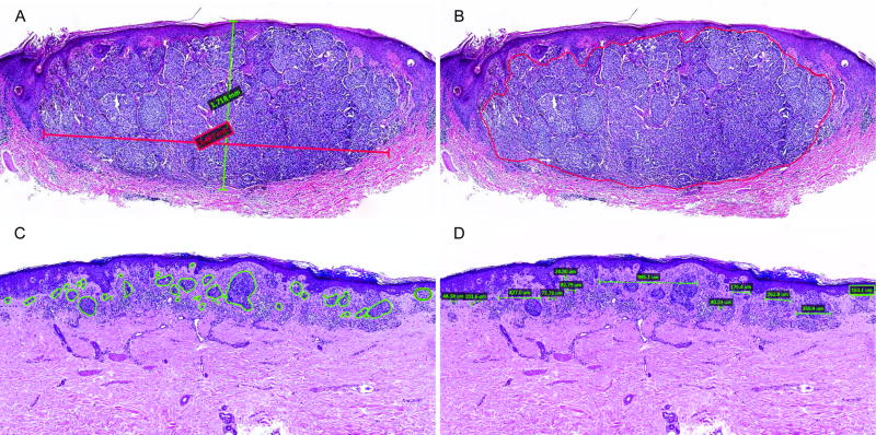
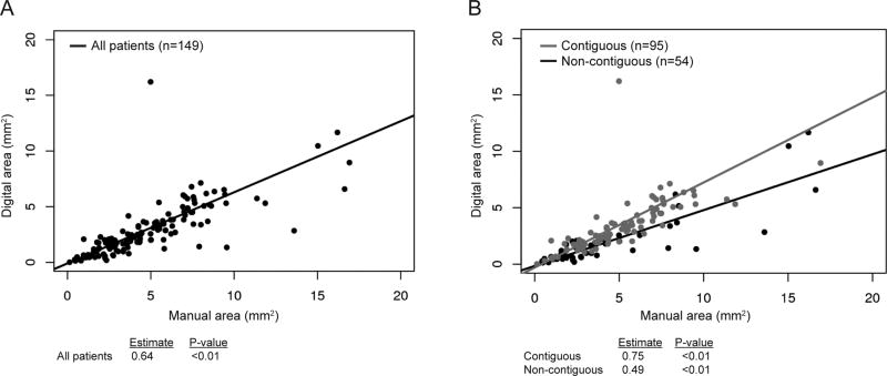
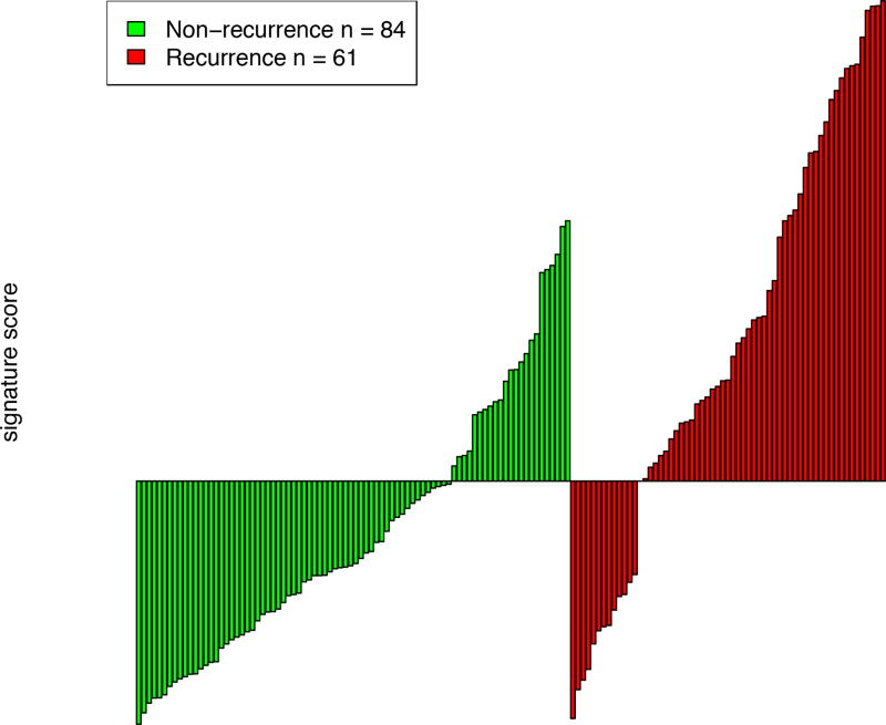
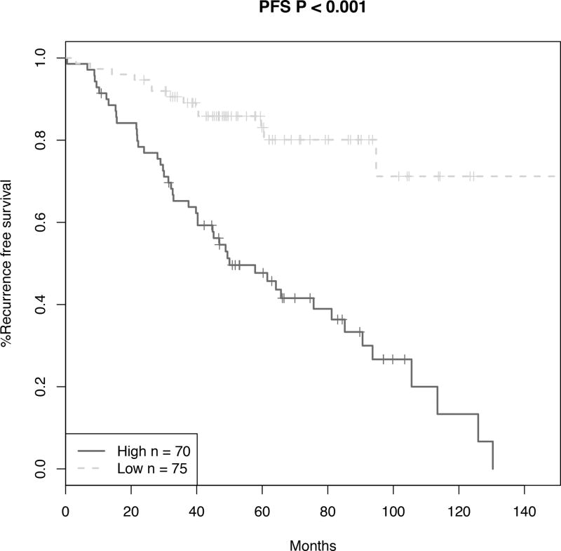
Current staging guidelines are insufficient to predict which patients with thin primary melanoma are at high risk of recurrence. Computer-assisted image analysis may allow for more practical and objective histopathological analysis of primary tumors than traditional light microscopy. We studied a prospective cohort of stage IB melanoma patients treated at NYU Langone Medical Center from 2002 to 2014. Primary tumor width, manual area, digital area, and conformation were evaluated in a patient subset via computer-assisted image analysis. The associations between histologic variables and survival were evaluated using Cox proportional hazards model. Logistic regressions were used to build a classifier with clinicopathological characteristics to predict recurrence status. Of the 655 patients with stage IB melanoma studied, a subset of 149 patient tumors (63 recurred, 86 did not recur) underwent computer-assisted histopathological analysis. Increasing tumor width (hazard ratios (HR): 1.17, P=0.01) and digital area (HR: 1.08, P<0.01) were significantly associated with worse recurrence-free survival, whereas non-contiguous conformation (HR: 0.57, P=0.05) was significantly associated with better recurrence-free survival. The novel histopathological classifier composed of digital area, conformation, and baseline variables effectively distinguished recurrent cases from non-recurrent cases (AUC: 0.733, 95% confidence interval (CI): 0.647-0.818), compared to the baseline classifier alone (AUC: 0.635, 95% CI: 0.545-0.724). Primary tumor cross-sectional area, width, and conformation measured via computer-assisted analysis may help identify high-risk patients with stage IB melanoma.
目前的分期指南不足以预测哪些原发性薄黑色素瘤患者有高复发风险。计算机辅助图像分析可能比传统的光学显微镜更能对原发性肿瘤进行更实用和客观的组织病理学分析。我们研究了一组 2002 年至 2014 年在纽约大学朗格尼医学中心治疗的 IB 期黑色素瘤患者的前瞻性队列。通过计算机辅助图像分析,对患者亚组的原发性肿瘤宽度、手动面积、数字面积和形态进行评估。使用 Cox 比例风险模型评估组织学变量与生存之间的关系。使用逻辑回归构建一个具有临床病理特征的分类器来预测复发状态。在研究的 655 例 IB 期黑色素瘤患者中,有 149 例患者的肿瘤(63 例复发,86 例未复发)进行了计算机辅助组织病理学分析。肿瘤宽度增加(危险比(HR):1.17,P=0.01)和数字面积(HR:1.08,P<0.01)与无复发生存率显著相关,而非连续形态(HR:0.57,P=0.05)与无复发生存率显著相关。由数字面积、形态和基线变量组成的新型组织病理学分类器能够有效区分复发病例和非复发病例(AUC:0.733,95%置信区间(CI):0.647-0.818),而与单独使用基线分类器相比(AUC:0.635,95%CI:0.545-0.724)。通过计算机辅助分析测量的原发性肿瘤横截面积、宽度和形态可能有助于识别 IB 期黑色素瘤的高危患者。