Prata Tiago S, Lopes Flavio S, Prado Vitor G, Almeida Izabela, Matsubara Igor, Dorairaj Syril, Furlanetto Rafael L, Vessani Roberto M, Paranhos Augusto
Glaucoma Service, Department of Ophthalmology, Federal University of São Paulo, São Paulo, Brazil.
Glaucoma Unit, Hospital Medicina dos Olhos, Osasco, Brazil.
PLoS One. 2017 Jul 21;12(7):e0180128. doi: 10.1371/journal.pone.0180128. eCollection 2017.
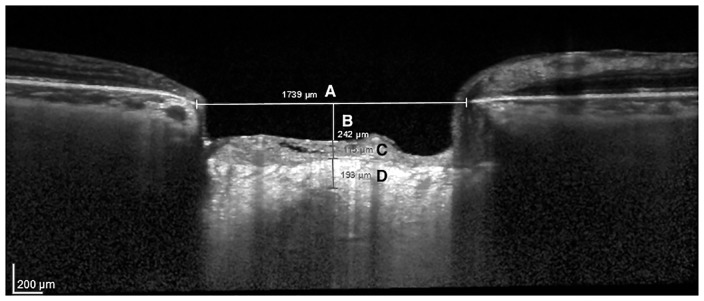
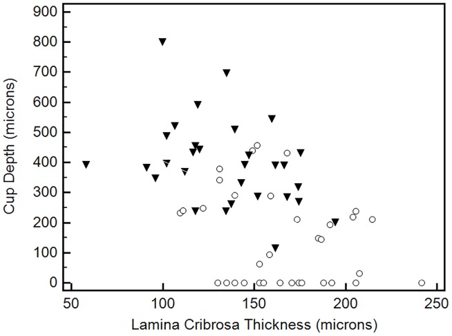
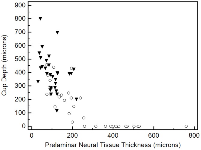
Structural differences between optic nerve head (ONH) parameters in glaucomatous and non-glaucomatous eyes has been documented, however the association between such parameters in patients with different disease stages is yet to be elucidated. We investigated the relationship between different laminar and prelaminar ONH structures using enhanced depth imaging spectral-domain optical coherence tomography (EDI OCT) in a population with and without glaucoma. In this observational case-control study, we prospectively enrolled healthy individuals and glaucomatous patients with different disease stages. All participants underwent EDI OCT imaging (Heidelberg Engineering). Following ONH parameters were measured on serial vertical B-scans by two examiners masked to patient's clinical data: lamina cribrosa (LC) and prelaminar neural tissue (PLNT) thicknesses, Bruch's membrane opening (BMO) and cup depth. Associations between cup depth, and laminar and prelaminar parameters were evaluated controlling for possible confounding factors such as axial length and disc size. Sixty-four eyes of 64 patients were included (30 with glaucoma and 34 controls). Eyes with glaucoma had significantly lower mean LC and PLNT thickness, and greater mean cup depth than controls (p<0.01). There was a significant negative association between PLNT thickness and cup depth in glaucomatous eyes (R2 = 0.158, p = 0.029). In addition, LC thickness correlated significantly with cup depth (R2 = 0.135, p = 0.045). Eyes with thinner LCs presented deeper cups. Overall, cup depth and BMO had the best and LC thickness had the worst intraobserver and interobserver reproducibility grading. In conclusion, significant associations were seen between cup depth, LC and PLNT thickness. Eyes with deeper cups not only had less neural tissue, but also thinner LCs, independent of disc size and axial length. Best reproducibility was found for prelaminar parameters compared to deeper ONH structures.
青光眼患者与非青光眼患者视神经乳头(ONH)参数的结构差异已有记录,但不同疾病阶段患者的这些参数之间的关联尚待阐明。我们使用增强深度成像光谱域光学相干断层扫描(EDI OCT)研究了有青光眼和无青光眼人群中不同板层和板层前ONH结构之间的关系。在这项观察性病例对照研究中,我们前瞻性纳入了健康个体和不同疾病阶段的青光眼患者。所有参与者均接受EDI OCT成像(海德堡工程公司)。两名对患者临床数据不知情的检查人员在连续垂直B扫描上测量以下ONH参数:筛板(LC)和板层前神经组织(PLNT)厚度、布鲁赫膜开口(BMO)和杯深。在控制可能的混杂因素(如眼轴长度和视盘大小)的情况下,评估杯深与板层和板层前参数之间的关联。纳入了64例患者的64只眼(30例青光眼患者和34例对照)。青光眼患者的平均LC和PLNT厚度显著低于对照组,平均杯深则显著大于对照组(p<0.01)。青光眼患者的PLNT厚度与杯深之间存在显著负相关(R2 = 0.158,p = 0.029)。此外,LC厚度与杯深显著相关(R2 = 0.135,p = 0.045)。LC较薄的眼杯更深。总体而言,杯深和BMO的观察者内和观察者间重复性分级最佳,而LC厚度最差。总之,杯深、LC和PLNT厚度之间存在显著关联。杯深较深的眼不仅神经组织较少,而且LC较薄,与视盘大小和眼轴长度无关。与更深的ONH结构相比,板层前参数的重复性最佳。