Institute for Lung Research, German Center for Lung Research, Universities of Giessen and Marburg Lung Centre, Philipps-University Marburg, 35043, Marburg, Germany.
Department for Cell Biology, Philipps-University Marburg, 35043, Marburg, Germany.
Sci Rep. 2017 Jul 24;7(1):6301. doi: 10.1038/s41598-017-06443-1.
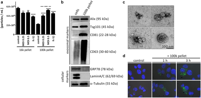
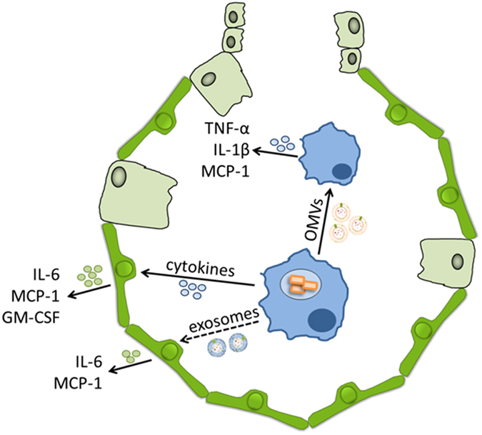
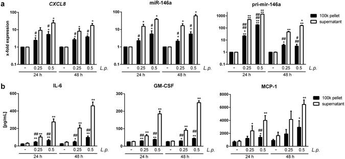
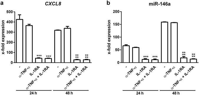
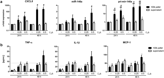
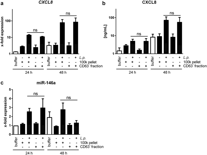
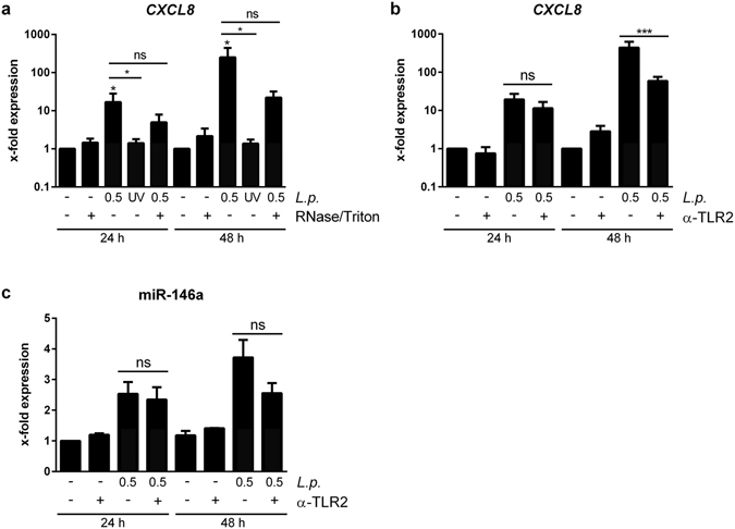
Extracellular vesicles from eukaryotic cells and outer membrane vesicles (OMVs) released from gram-negative bacteria have been described as mediators of pathogen-host interaction and intercellular communication. Legionella pneumophila (L. pneumophila) is a causative agent of severe pneumonia. The differential effect of bacterial and host cell vesicles in L. pneumophila infection is unknown so far. We infected THP-1-derived or primary human macrophages with L. pneumophila and isolated supernatant vesicles by differential centrifugation. We observed an increase of exosomes in the 100 k pellet by nanoparticle tracking analysis, electron microscopy, and protein markers. This fraction additionally contained Legionella LPS, indicating also the presence of OMVs. In contrast, vesicles in the 16 k pellet, representing microparticles, decreased during infection. The 100 k vesicle fraction activated uninfected primary human alveolar epithelial cells, A549 cells, and THP-1 cells. Epithelial cell activation was reduced by exosome depletion (anti-CD63, or GW4869), or blocking of IL-1β in the supernatant. In contrast, the response of THP-1 cells to vesicles was reduced by a TLR2-neutralizing antibody, UV-inactivation of bacteria, or - partially - RNase-treatment of vesicles. Taken together, we found that during L. pneumophila infection, neighbouring epithelial cells were predominantly activated by exosomes and cytokines, whereas myeloid cells were activated by bacterial OMVs.
真核细胞来源的细胞外囊泡和革兰氏阴性菌释放的外膜囊泡(OMVs)已被描述为病原体-宿主相互作用和细胞间通讯的介质。嗜肺军团菌(L. pneumophila)是严重肺炎的病原体。迄今为止,细菌和宿主细胞囊泡在 L. pneumophila 感染中的差异效应尚不清楚。我们用 L. pneumophila 感染 THP-1 衍生或原代人巨噬细胞,并通过差速离心分离上清囊泡。我们通过纳米颗粒跟踪分析、电子显微镜和蛋白质标志物观察到 100k 沉淀中外泌体增加。该部分还含有军团菌 LPS,表明也存在 OMVs。相比之下,感染过程中 16k 沉淀中的囊泡(代表微粒)减少。100k 囊泡部分激活未感染的原代人肺泡上皮细胞、A549 细胞和 THP-1 细胞。上清液中外泌体耗竭(抗 CD63 或 GW4869)或阻断 IL-1β 可降低上皮细胞的激活。相反,THP-1 细胞对囊泡的反应被 TLR2 中和抗体、细菌 UV 失活或部分 RNA 酶处理的囊泡降低。总之,我们发现,在 L. pneumophila 感染期间,邻近的上皮细胞主要被外泌体和细胞因子激活,而髓样细胞被细菌 OMVs 激活。