Milosevic Zorica C, Nadrljanski Mirjan M, Milovanovic Zorka M, Gusic Nina Z, Vucicevic Slavko S, Radulovic Olga S
Clinic for Radiation Oncology and Radiology, Institute of Oncology and Radiology of Serbia, Belgrade, Serbia.
Faculty of Medicine, University of Belgrade, Belgrade, Serbia.
Radiol Oncol. 2017 May 7;51(2):130-136. doi: 10.1515/raon-2017-0016. eCollection 2017 Jun.
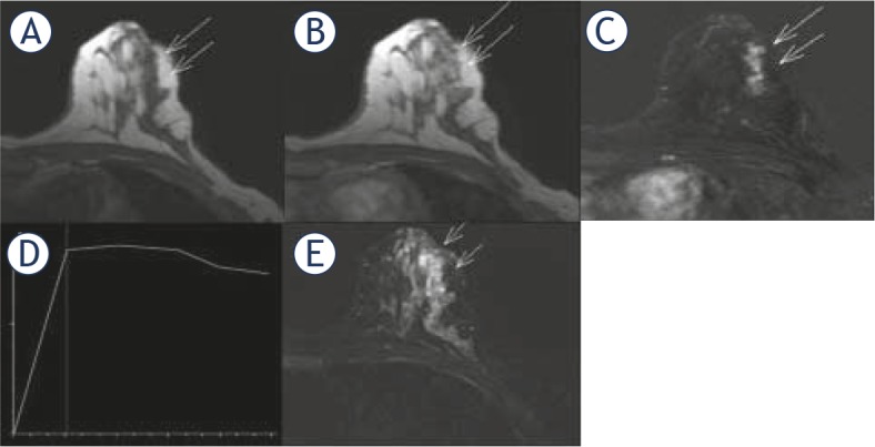
We aimed to analyse the morphokinetic features of breast fibrocystic changes (nonproliferative lesions, proliferative lesions without atypia and proliferative lesions with atypia) presenting as a non-mass enhancement (NME)in dynamic contrast-enhanced magnetic resonance imaging (DCE-MRI) examination.
Forty-six patients with histologically proven fibrocystic changes (FCCs) were retrospectively reviewed, according to Breast Imaging Reporting and Data System (BI-RADS) lexicon. Prior to DCE-MRI examination, a unilateral breast lesion suspicious of malignancy was detected clinically, on mammography or breast ultrasonography.
The predominant features of FCCs presenting as NME in DCE-MRI examination were: unilateral regional or diffuse distribution (in 35 patients or 76.1%), heterogeneous or clumped internal pattern of enhancement (in 36 patients or 78.3%), plateau time-intensity curve (in 25 patients or 54.3%), moderate or fast wash-in (in 31 patients or 67.4%).Nonproliferative lesions were found in 11 patients (24%), proliferative lesions without atypia in 29 patients (63%) and lesions with atypia in six patients (13%), without statistically significant difference of morphokinetic features, except of the association of clustered microcysts with proliferative dysplasia without atypia.
FCCs presenting as NME in DCE-MRI examination have several morphokinetic features suspicious of malignancy, therefore requiring biopsy (BI-RADS 4). Nonproliferative lesions, proliferative lesions without atypia and proliferative lesions with atypia predominantly share the same predefined DCE-MRI morphokinetic features.
我们旨在分析在动态对比增强磁共振成像(DCE-MRI)检查中表现为非肿块强化(NME)的乳腺纤维囊性变(非增殖性病变、无异型增生的增殖性病变和有异型增生的增殖性病变)的形态动力学特征。
根据乳腺影像报告和数据系统(BI-RADS)词典,对46例经组织学证实为纤维囊性变(FCCs)的患者进行回顾性分析。在DCE-MRI检查前,通过乳腺钼靶或乳腺超声在临床上检测到单侧乳腺可疑恶性病变。
在DCE-MRI检查中表现为NME的FCCs的主要特征为:单侧区域性或弥漫性分布(35例患者,占76.1%)、内部强化不均匀或呈团块状(36例患者,占78.3%)、平台期时间-强度曲线(25例患者,占54.3%)、中等或快速流入(31例患者,占67.4%)。11例患者(24%)为非增殖性病变,29例患者(63%)为无异型增生的增殖性病变,6例患者(13%)为有异型增生的病变,除了簇状微囊肿与无异型增生的增殖性发育异常有关外,形态动力学特征无统计学显著差异。
在DCE-MRI检查中表现为NME的FCCs具有一些可疑恶性的形态动力学特征,因此需要活检(BI-RADS 4类)。非增殖性病变、无异型增生的增殖性病变和有异型增生的增殖性病变主要具有相同的预定义DCE-MRI形态动力学特征。