• 文献检索
  • 文档翻译
  • 深度研究
  • 学术资讯
  • Suppr Zotero 插件Zotero 插件
  • 邀请有礼
  • 套餐&价格
  • 历史记录
应用&插件
Suppr Zotero 插件Zotero 插件浏览器插件Mac 客户端Windows 客户端微信小程序
定价
高级版会员购买积分包购买API积分包
服务
文献检索文档翻译深度研究API 文档MCP 服务
关于我们
关于 Suppr公司介绍联系我们用户协议隐私条款
关注我们

Suppr 超能文献

核心技术专利:CN118964589B侵权必究
粤ICP备2023148730 号-1Suppr @ 2026

文献检索

告别复杂PubMed语法,用中文像聊天一样搜索,搜遍4000万医学文献。AI智能推荐,让科研检索更轻松。

立即免费搜索

文件翻译

保留排版,准确专业,支持PDF/Word/PPT等文件格式,支持 12+语言互译。

免费翻译文档

深度研究

AI帮你快速写综述,25分钟生成高质量综述,智能提取关键信息,辅助科研写作。

立即免费体验

基于乳腺 MRI 的纹理分析在鉴别绝经前妇女良性和恶性非肿块样强化中的附加价值。

Additive value of texture analysis based on breast MRI for distinguishing between benign and malignant non-mass enhancement in premenopausal women.

机构信息

Department of Radiology, Guangdong Women and Children Hospital, No.521, Xingnan Road, Panyu District, Guangzhou, 511400, China.

Department of Radiology, The Third Affiliated Hospital of Guangzhou Medical University, Guangzhou, China.

出版信息

BMC Med Imaging. 2021 Mar 12;21(1):48. doi: 10.1186/s12880-021-00571-x.

DOI:10.1186/s12880-021-00571-x
PMID:33706695
原文链接:https://pmc.ncbi.nlm.nih.gov/articles/PMC7953679/
Abstract

BACKGROUND

Non-mass enhancement (NME) is a diagnostic dilemma and highly reliant on the experience of the radiologists. Texture analysis (TA) could serve as an objective method to quantify lesion characteristics. However, it remains unclear what role TA plays in a predictive model based on routine MRI characteristics. The purpose of this study was to explore the value of TA in distinguishing between benign and malignant NME in premenopausal women.

METHODS

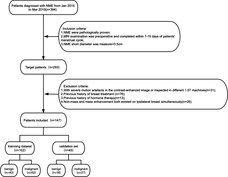
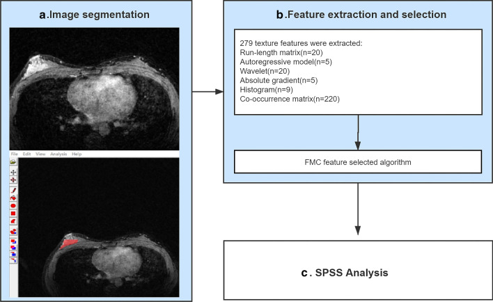
Women in whom NME was histologically proven (n = 147) were enrolled (benign: 58; malignant: 89) was retrospective. Then, 102 and 45 patients were classified as the training and validation groups, respectively. Scanning sequences included Fat-suppressed T2-weighted and fat-suppressed contrast-enhanced T1-weighted which were acquired on a 1.5T MRI system. Clinical and routine MR characteristics (CRMC) were evaluated by two radiologists according to the Breast Imaging and Reporting and Data system (2013). Texture features were extracted from all post-contrast sequences in the training group. The combination model was built and then assessed in the validation group. Pearson's chi-square test and Mann-Whitney U test were used to compare categorical variables and continuous variables, respectively. Logistic regression analysis and receiver operating characteristic curve were employed to assess the diagnostic performance of CRMC, TA, and their combination model in NME diagnosis.

RESULTS

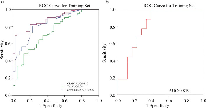
The combination model showed superior diagnostic performance in differentiating between benign and malignant NME compared to that of CRMC or TA alone (AUC, 0.887 vs 0.832 vs 0.74). Moreover, compared to CRMC, the model showed high specificity (72.5% vs 80%). The results obtained in the validation group confirmed the model was promising.

CONCLUSIONS

With the combined use of TA and CRMC could afford an improved diagnostic performance in differentiating between benign and malignant NME.

摘要

背景

非肿块样强化(NME)是一种诊断难题,高度依赖于放射科医生的经验。纹理分析(TA)可以作为量化病变特征的客观方法。然而,基于常规 MRI 特征的预测模型中 TA 扮演什么角色仍不清楚。本研究旨在探讨 TA 在鉴别绝经前妇女良恶性 NME 中的作用。

方法

回顾性纳入经组织学证实的 NME 患者(良性:58 例;恶性:89 例)。然后,将 102 例和 45 例患者分别分为训练组和验证组。扫描序列包括脂肪抑制 T2 加权和脂肪抑制对比增强 T1 加权,均在 1.5T MRI 系统上采集。两名放射科医生根据乳腺影像报告和数据系统(2013 年)评估临床和常规磁共振特征(CRMC)。在训练组中,从所有增强后序列中提取纹理特征。建立组合模型,然后在验证组中进行评估。采用 Pearson χ2 检验和 Mann-Whitney U 检验分别比较分类变量和连续变量。采用逻辑回归分析和受试者工作特征曲线评估 CRMC、TA 及其组合模型在 NME 诊断中的诊断性能。

结果

与 CRMC 或 TA 单独相比,组合模型在鉴别良恶性 NME 方面表现出更好的诊断性能(AUC:0.887 比 0.832 比 0.74)。此外,与 CRMC 相比,该模型具有较高的特异性(72.5%比 80%)。验证组的结果证实了该模型的前景。

结论

TA 和 CRMC 的联合使用可以提高鉴别良恶性 NME 的诊断性能。

https://cdn.ncbi.nlm.nih.gov/pmc/blobs/23b2/7953679/b76df4387b7d/12880_2021_571_Fig4_HTML.jpg
https://cdn.ncbi.nlm.nih.gov/pmc/blobs/23b2/7953679/e14ada53a2b1/12880_2021_571_Fig1_HTML.jpg
https://cdn.ncbi.nlm.nih.gov/pmc/blobs/23b2/7953679/53ebcf717e88/12880_2021_571_Fig2_HTML.jpg
https://cdn.ncbi.nlm.nih.gov/pmc/blobs/23b2/7953679/bad795510726/12880_2021_571_Fig3_HTML.jpg
https://cdn.ncbi.nlm.nih.gov/pmc/blobs/23b2/7953679/b76df4387b7d/12880_2021_571_Fig4_HTML.jpg

相似文献

1
Additive value of texture analysis based on breast MRI for distinguishing between benign and malignant non-mass enhancement in premenopausal women.基于乳腺 MRI 的纹理分析在鉴别绝经前妇女良性和恶性非肿块样强化中的附加价值。
BMC Med Imaging. 2021 Mar 12;21(1):48. doi: 10.1186/s12880-021-00571-x.
2
Contrasts Between Diffusion-Weighted Imaging and Dynamic Contrast-Enhanced MR in Diagnosing Malignancies of Breast Nonmass Enhancement Lesions Based on Morphologic Assessment.基于形态学评估的扩散加权成像与动态对比增强磁共振在诊断乳腺非肿块样强化病变恶性肿瘤中的对比。
J Magn Reson Imaging. 2023 Sep;58(3):963-974. doi: 10.1002/jmri.28600. Epub 2023 Feb 3.
3
Diagnostic value of mammography for accompanying non-mass enhancement on preoperative breast MRI.术前乳腺 MRI 伴随非肿块样强化的乳腺 X 线摄影诊断价值。
Acta Radiol. 2022 Aug;63(8):1032-1042. doi: 10.1177/02841851211030771. Epub 2021 Jul 14.
4
The diagnostic value of multimodal imaging based on MR combined with ultrasound in benign and malignant breast diseases.基于 MR 联合超声的多模态成像在良恶性乳腺疾病中的诊断价值。
Clin Exp Med. 2024 May 23;24(1):110. doi: 10.1007/s10238-024-01377-1.
5
Value of conventional magnetic resonance imaging texture analysis in the differential diagnosis of benign and borderline/malignant phyllodes tumors of the breast.常规磁共振成像纹理分析在鉴别诊断乳腺良性和交界性/恶性叶状肿瘤中的价值。
Cancer Imaging. 2021 Mar 12;21(1):29. doi: 10.1186/s40644-021-00398-3.
6
Magnetic resonance imaging features for differentiating breast papilloma with high-risk or malignant lesions from benign papilloma: a retrospective study on 158 patients.磁共振成像特征鉴别高危或恶性病变乳腺乳头状瘤与良性乳头状瘤:158 例回顾性研究。
World J Surg Oncol. 2018 Dec 17;16(1):234. doi: 10.1186/s12957-018-1537-9.
7
Performance evaluation of texture analysis based on kinetic parametric maps from breast DCE-MRI in classifying benign from malignant lesions.基于乳腺 DCE-MRI 动力学参数图的纹理分析在鉴别良恶性病变中的性能评估。
J Surg Oncol. 2020 Jun;121(8):1181-1190. doi: 10.1002/jso.25901. Epub 2020 Mar 13.
8
An artificial intelligence system using maximum intensity projection MR images facilitates classification of non-mass enhancement breast lesions.基于最大密度投影磁共振图像的人工智能系统有助于非肿块样强化乳腺病变的分类。
Eur Radiol. 2022 Jul;32(7):4857-4867. doi: 10.1007/s00330-022-08553-5. Epub 2022 Mar 8.
9
Limited role of DWI with apparent diffusion coefficient mapping in breast lesions presenting as non-mass enhancement on dynamic contrast-enhanced MRI.弥散加权成像及其表观扩散系数图在动态对比增强 MRI 呈非肿块样强化的乳腺病变中的作用有限。
Breast Cancer Res. 2019 Dec 4;21(1):136. doi: 10.1186/s13058-019-1208-y.
10
Investigating the diagnostic value of quantitative parameters based on T2-weighted and contrast-enhanced MRI with psoas muscle and outer myometrium as internal references for differentiating uterine sarcomas from leiomyomas at 3T MRI.探讨以腰大肌和子宫外肌作为内参照在 3T MRI 下基于 T2 加权和对比增强 MRI 的定量参数对子宫肉瘤与子宫肌瘤的鉴别诊断价值。
Cancer Imaging. 2019 Apr 1;19(1):20. doi: 10.1186/s40644-019-0206-8.

引用本文的文献

1
The value of intratumoral and peritumoral ultrasound radiomics model constructed using multiple machine learning algorithms for non-mass breast cancer.使用多种机器学习算法构建的瘤内和瘤周超声影像组学模型对非肿块型乳腺癌的价值。
Sci Rep. 2025 Jun 6;15(1):19953. doi: 10.1038/s41598-025-03704-2.
2
Ultrasound diagnosis of non-mass MRI-detected lesions.MRI 检测阴性非肿块性病变的超声诊断。
J Med Ultrason (2001). 2023 Jul;50(3):351-360. doi: 10.1007/s10396-023-01306-x. Epub 2023 Apr 29.
3
Radiomics in breast MRI: current progress toward clinical application in the era of artificial intelligence.

本文引用的文献

1
Diffusion-weighted imaging or dynamic contrast-enhanced curve: a retrospective analysis of contrast-enhanced magnetic resonance imaging-based differential diagnoses of benign and malignant breast lesions.弥散加权成像或动态对比增强曲线:基于对比增强磁共振成像的良性和恶性乳腺病变鉴别诊断的回顾性分析。
Eur Radiol. 2020 Sep;30(9):4795-4805. doi: 10.1007/s00330-020-06883-w. Epub 2020 Apr 29.
2
Texture Analysis with 3.0-T MRI for Association of Response to Neoadjuvant Chemotherapy in Breast Cancer.基于 3.0T MRI 的纹理分析对乳腺癌新辅助化疗反应的相关性研究。
Radiology. 2020 Jan;294(1):31-41. doi: 10.1148/radiol.2019182718. Epub 2019 Nov 26.
3
乳腺 MRI 的放射组学:人工智能时代向临床应用的当前进展。
Radiol Med. 2022 Jan;127(1):39-56. doi: 10.1007/s11547-021-01423-y. Epub 2021 Oct 26.
4
Non-Mass Enhancements on DCE-MRI: Development and Validation of a Radiomics-Based Signature for Breast Cancer Diagnoses.动态对比增强磁共振成像中的非肿块强化:基于影像组学的乳腺癌诊断特征的开发与验证
Front Oncol. 2021 Sep 22;11:738330. doi: 10.3389/fonc.2021.738330. eCollection 2021.
The Utility of Texture Analysis Based on Breast Magnetic Resonance Imaging in Differentiating Phyllodes Tumors From Fibroadenomas.
基于乳腺磁共振成像的纹理分析在鉴别叶状肿瘤与纤维腺瘤中的应用
Front Oncol. 2019 Oct 15;9:1021. doi: 10.3389/fonc.2019.01021. eCollection 2019.
4
Contrast-Enhanced Mammography and Radiomics Analysis for Noninvasive Breast Cancer Characterization: Initial Results.对比增强乳腺摄影和放射组学分析在无创性乳腺癌特征描述中的应用:初步结果。
Mol Imaging Biol. 2020 Jun;22(3):780-787. doi: 10.1007/s11307-019-01423-5.
5
Role of texture analysis in breast MRI as a cancer biomarker: A review.纹理分析在乳腺 MRI 作为癌症生物标志物中的作用:综述。
J Magn Reson Imaging. 2019 Apr;49(4):927-938. doi: 10.1002/jmri.26556. Epub 2018 Nov 3.
6
Diagnostic performance of initial enhancement analysis using ultra-fast dynamic contrast-enhanced MRI for breast lesions.使用超快速动态对比增强 MRI 对乳腺病变进行初始增强分析的诊断性能。
Eur Radiol. 2019 Mar;29(3):1164-1174. doi: 10.1007/s00330-018-5643-4. Epub 2018 Aug 7.
7
Precision Medicine and Radiogenomics in Breast Cancer: New Approaches toward Diagnosis and Treatment.精准医学与乳腺癌放射组学:诊断与治疗的新方法。
Radiology. 2018 Jun;287(3):732-747. doi: 10.1148/radiol.2018172171.
8
MRI background parenchymal enhancement, breast density and serum hormones in postmenopausal women.MRI 脑实质增强、绝经后妇女的乳房密度和血清激素。
Int J Cancer. 2018 Aug 15;143(4):823-830. doi: 10.1002/ijc.31370. Epub 2018 Mar 25.
9
Revisiting Nonmass Enhancement in Breast MRI: Analysis of Outcomes and Follow-Up Using the Updated BI-RADS Atlas.重新审视乳腺MRI中的非肿块强化:使用更新版BI-RADS图谱分析结果及随访情况。
AJR Am J Roentgenol. 2017 Nov;209(5):1178-1184. doi: 10.2214/AJR.17.18086. Epub 2017 Aug 23.
10
MRI Texture Analysis of Background Parenchymal Enhancement of the Breast.乳腺背景实质强化的MRI纹理分析
Biomed Res Int. 2017;2017:4845909. doi: 10.1155/2017/4845909. Epub 2017 Jul 24.