Shoji Takuhei, Kuroda Hiroto, Suzuki Masayuki, Ibuki Hisashi, Araie Makoto, Yoneya Shin
Department of Ophthalmology, Saitama Medical University, Iruma, Saitama, Japan.
Advanced Laser Medical Center, Department of Ophthalmology, Saitama Medical University, Iruma, Saitama, Japan.
PLoS One. 2017 Jul 25;12(7):e0181675. doi: 10.1371/journal.pone.0181675. eCollection 2017.
The lamina cribrosa (LC) is known to play a critical role in the pathogenesis of glaucoma. Although it has been reported that striae-shaped or slit-shaped lamina pores are more frequent in eyes with primary open angle glaucoma (POAG), this observation is based only on fundus photography. The primary object of this study is to perform layer-by-layer comparisons of the shape of lamina pores within the LC in vivo.
Cross-sectional study.
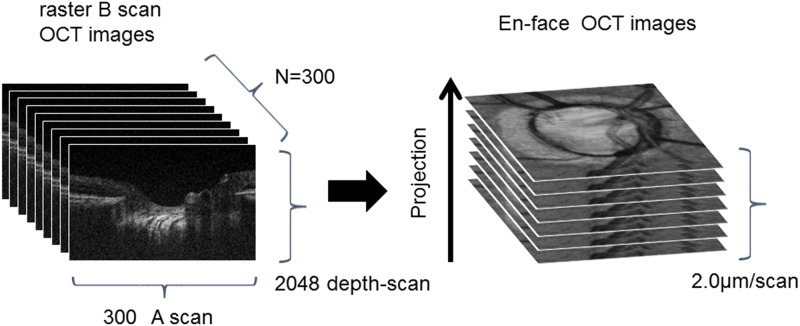
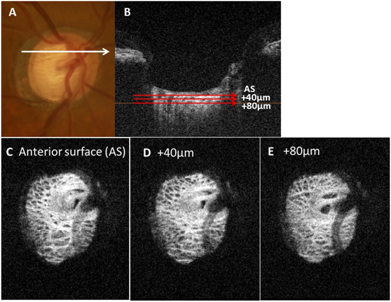
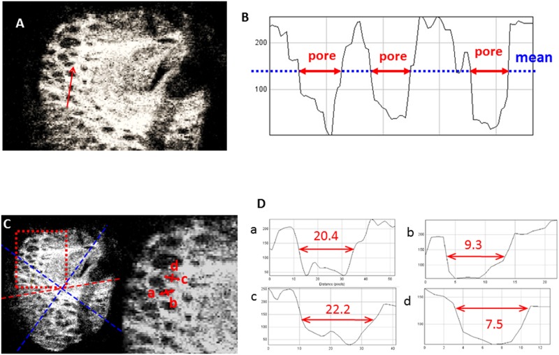
Optic nerve head B-scans were obtained using custom-made broad-wavelength optical coherence tomography with a mode-locked laser. A total of 300 single B-scans per eye were obtained and three-dimensional images were rendered from these image sequences to obtain 2-μm thin-slice en face images of the LC. Elongation indices (EIs) of the lamina pores were measured from the anterior surface (AS) of the LC to the deeper layers in 40-μm increments.
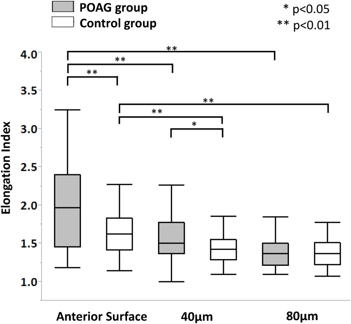
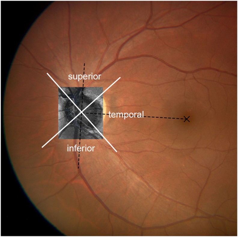
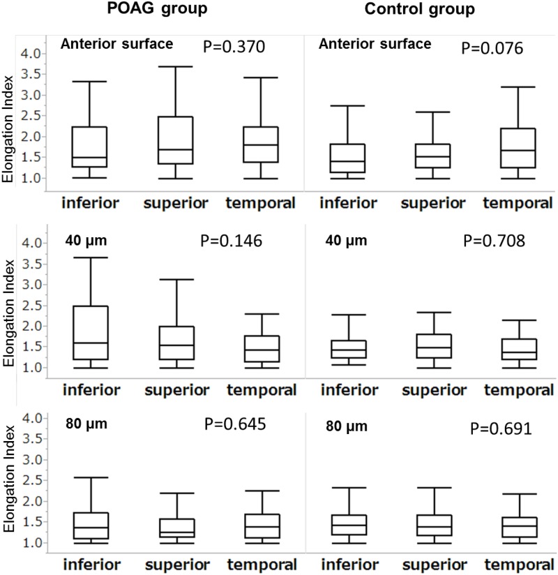
Thirteen eyes from 10 primary open angle glaucoma (POAG) patients of mean deviation -15.2 (-16.5, -12.9) (median [25,75 percentile]) dB and 10 eyes from 7 normal controls were studied. Although the EI value was not significantly different between the superior, temporal and inferior regions of the LC at any depth level in either group, it was greater at the AS than at the 40 μm and 80 μm depth levels (P < .001) in both groups, and was greater in the POAG group only at the AS and 40 μm depth level (P ≤ .05). After adjustment for age and refraction, the effects of depth and presence of POAG on the EI value remained significant. Also, the severity of glaucoma and depth were significant factors associated with EI in multivariate analysis.
Elongation of lamina pores was significantly more evident at the anterior surface and the 40-μm depth level of the LC in POAG eyes than in normal eyes, suggesting that nerve fiber bundles passing through the LC were under greater stress in the anterior layers of the LC.
已知筛板(LC)在青光眼发病机制中起关键作用。尽管已有报道称,在原发性开角型青光眼(POAG)患者眼中,条纹状或裂隙状筛板孔更为常见,但该观察仅基于眼底照相。本研究的主要目的是在体内对LC内筛板孔的形状进行逐层比较。
横断面研究。
使用定制的宽波长光学相干断层扫描结合锁模激光获取视神经乳头B扫描图像。每只眼睛共获得300幅单幅B扫描图像,并从这些图像序列生成三维图像,以获得LC的2μm薄层正面图像。从LC的前表面(AS)开始,以40μm的增量测量筛板孔的伸长指数(EI)至更深层。
研究了10例原发性开角型青光眼(POAG)患者的13只眼睛,平均偏差为-15.2(-16.5,-12.9)(中位数[第25,75百分位数])dB,以及7例正常对照者的10只眼睛。尽管在任何深度水平上,两组中LC的上、颞和下区域之间的EI值均无显著差异,但两组中AS处的EI值均大于40μm和80μm深度水平处(P <.001),且仅在POAG组中,AS和40μm深度水平处的EI值更大(P≤.05)。在调整年龄和屈光度后,深度和POAG的存在对EI值的影响仍然显著。此外,在多变量分析中,青光眼的严重程度和深度是与EI相关的显著因素。
与正常眼睛相比,POAG眼睛中筛板孔的伸长在LC的前表面和40μm深度水平处明显更明显,这表明穿过LC的神经纤维束在LC的前层承受更大的压力。