Zhou Shukui, Yang Ranxin, Zou Qingsong, Zhang Kaile, Yin Ting, Zhao Weixin, Shapter Joseph G, Gao Guo, Fu Qiang
Department of Urology, Affiliated Sixth People's Hospital, Shanghai Jiao Tong University, Shanghai, China.
Institute of Nano Biomedicine and Engineering, Department of Instrument Science and Technology, School of Electronic Information and Electrical Engineering, Shanghai Jiao Tong University, Shanghai, China.
Theranostics. 2017 Jun 25;7(9):2509-2523. doi: 10.7150/thno.18833. eCollection 2017.
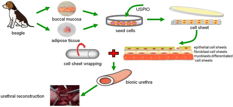
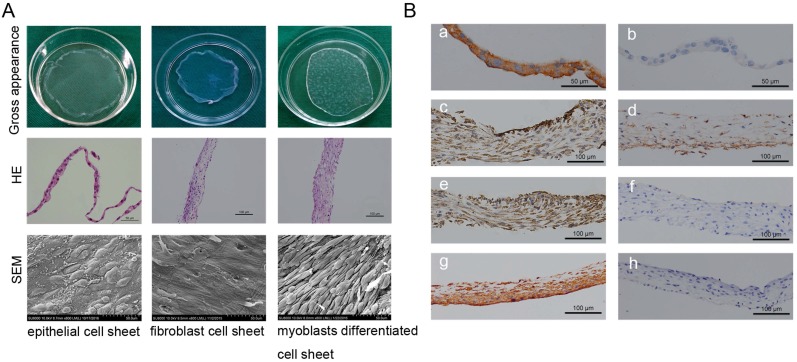
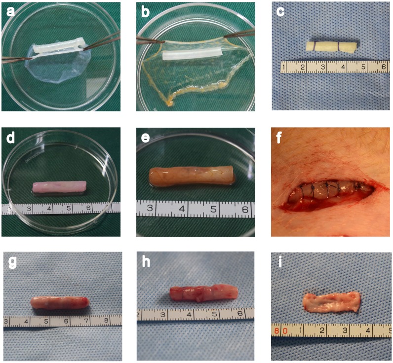
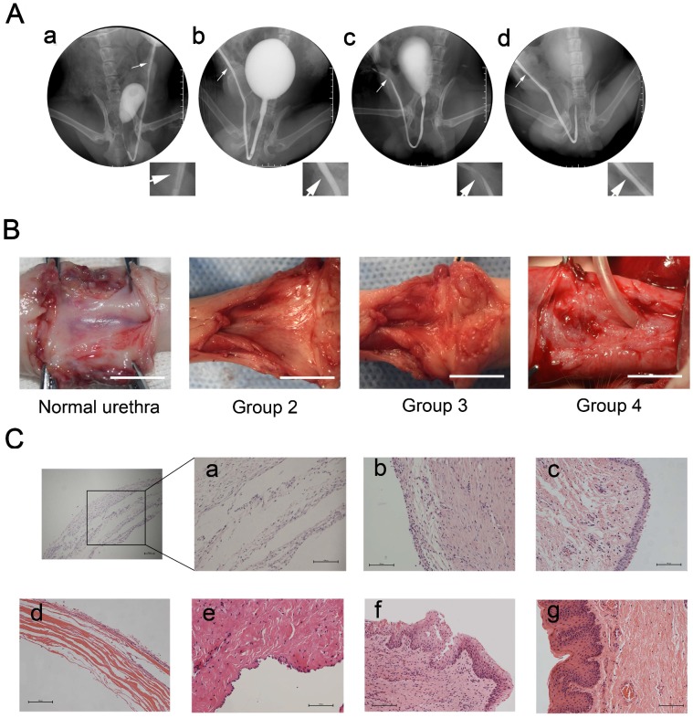
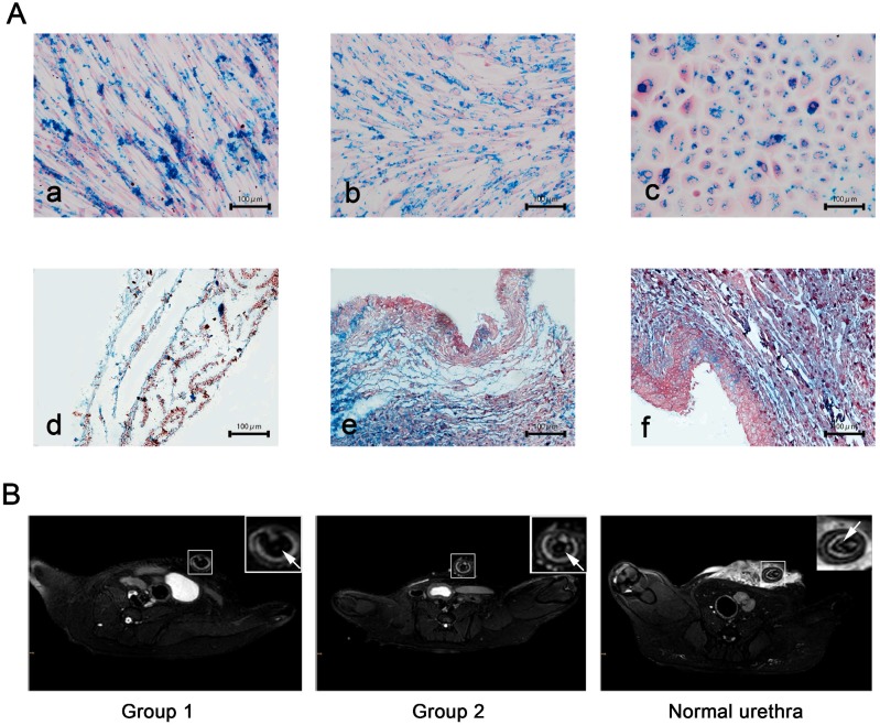
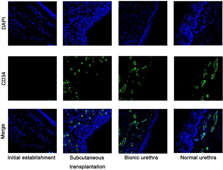
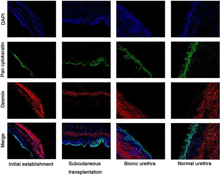
Urethral strictures remain a reconstructive challenge, due to less than satisfactory outcomes and high incidence of stricture recurrence. An "ideal" urethral reconstruction should establish similar architecture and function as the original urethral wall. We fabricated a novel tissue-engineered bionic urethras using cell sheet technology and report their viability in a canine model. Small amounts of oral and adipose tissues were harvested, and adipose-derived stem cells, oral mucosal epithelial cells, and oral mucosal fibroblasts were isolated and used to prepare cell sheets. The cell sheets were hierarchically tubularized to form 3-layer tissue-engineered urethras and labeled by ultrasmall super-paramagnetic iron oxide (USPIO). The constructed tissue-engineered urethras were transplanted subcutaneously for 3 weeks to promote the revascularization and biomechanical strength of the implant. Then, 2 cm length of the tubularized penile urethra was replaced by tissue-engineered bionic urethra. At 3 months of urethral replacement, USPIO-labeled tissue-engineered bionic urethra can be effectively detected by MRI at the transplant site. Histologically, the retrieved bionic urethras still displayed 3 layers, including an epithelial layer, a fibrous layer, and a myoblast layer. Three weeks after subcutaneous transplantation, immunofluorescence analysis showed the density of blood vessels in bionic urethra was significantly increased following the initial establishment of the constructs and was further up-regulated at 3 months after urethral replacement and was close to normal level in urethral tissue. Our study is the first to experimentally demonstrate 3-layer tissue-engineered urethras can be established using cell sheet technology and can promote the regeneration of structural and functional urethras similar to normal urethra.
由于治疗效果不尽人意以及狭窄复发率高,尿道狭窄仍然是一个重建方面的挑战。“理想的”尿道重建应建立与原始尿道壁相似的结构和功能。我们使用细胞片技术制造了一种新型的组织工程化仿生尿道,并报告了它们在犬模型中的生存能力。采集少量口腔和脂肪组织,分离脂肪来源的干细胞、口腔黏膜上皮细胞和口腔黏膜成纤维细胞并用于制备细胞片。将细胞片分层管状化以形成三层组织工程化尿道,并用超小超顺磁性氧化铁(USPIO)进行标记。将构建好的组织工程化尿道皮下移植3周,以促进植入物的血管再生和生物力学强度。然后,用组织工程化仿生尿道替换2厘米长的管状阴茎尿道。在尿道置换3个月时,MRI能够在移植部位有效检测到USPIO标记的组织工程化仿生尿道。组织学检查显示,取回的仿生尿道仍呈现三层结构,包括上皮层、纤维层和平滑肌层。皮下移植3周后,免疫荧光分析显示,构建物初步形成后,仿生尿道中的血管密度显著增加,在尿道置换3个月时进一步上调,接近尿道组织中的正常水平。我们的研究首次通过实验证明,使用细胞片技术可以构建三层组织工程化尿道,并能促进类似于正常尿道的结构和功能尿道的再生。