Alkhalil Mohammad, Biasiolli Luca, Chai Joshua T, Galassi Francesca, Li Linqing, Darby Christopher, Halliday Alison, Hands Linda, Magee Timothy, Perkins Jeremy, Sideso Ed, Jezzard Peter, Robson Matthew D, Handa Ashok, Choudhury Robin P
Acute Vascular Imaging Centre, Radcliffe Department of Medicine, University of Oxford, Oxford, United Kingdom.
FMRIB Centre, Nuffield Department of Clinical Neurosciences, University of Oxford, Oxford, United Kingdom.
PLoS One. 2017 Jul 26;12(7):e0181668. doi: 10.1371/journal.pone.0181668. eCollection 2017.
Techniques to stratify subgroups of patients with asymptomatic carotid artery disease are urgently needed to guide decisions on optimal treatment. Reliance on estimates of % luminal stenosis has not been effective, perhaps because that approach entirely disregards potentially important information on the pathological process in the wall of the artery.
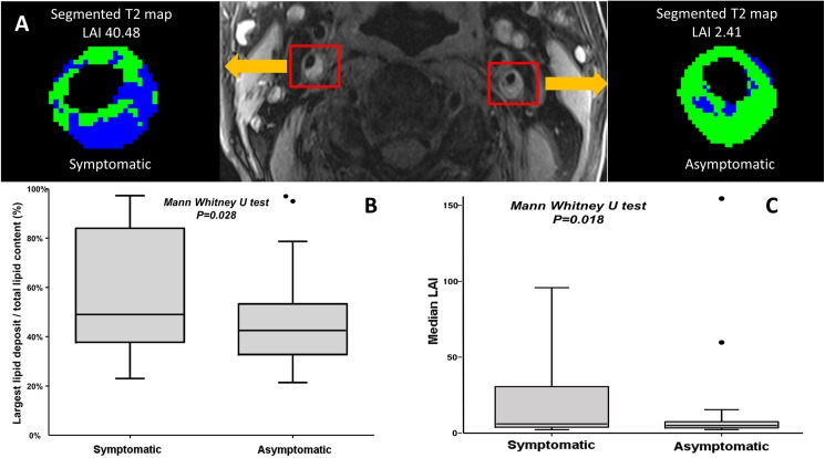
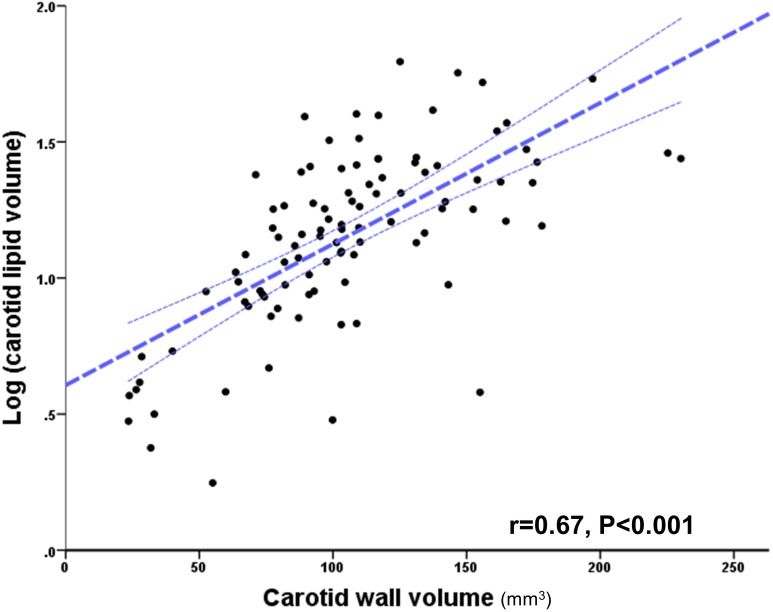
Since plaque lipid is a key determinant of plaque behaviour we used a newly validated, high-sensitivity T2-mapping MR technique for a systematic survey of the quantity and distribution of plaque lipid in patients undergoing endarterectomy. Lipid percentage was quantified in 50 carotid endarterectomy patients. Lipid distribution was tested, using two imaging indices (contribution of the largest lipid deposit towards total lipid (LLD %) and a newly-developed LAI 'lipid aggregation index').
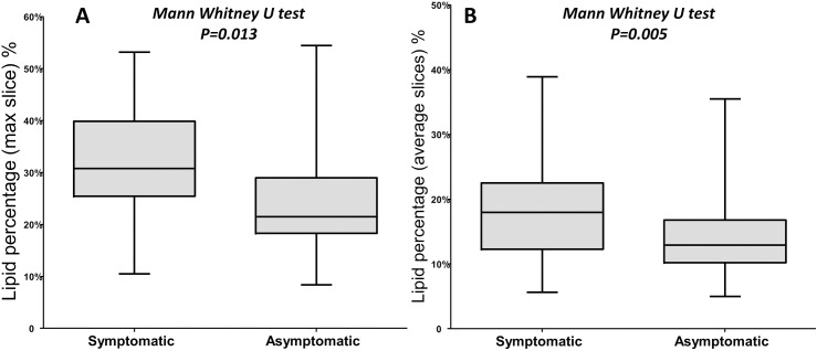
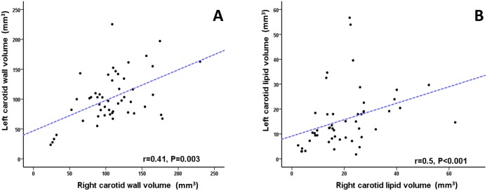
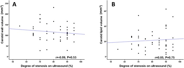
The bifurcation contained maximal lipid volume. Lipid percentage was higher in symptomatic vs. asymptomatic patients with degree of stenosis (DS ≥ 50%) and in the total cohort (P = 0.013 and P = 0.005, respectively). Both LLD % and LAI was higher in symptomatic patients (P = 0.028 and P = 0.018, respectively), suggesting that for a given plaque lipid volume, coalesced deposits were more likely to be associated with symptomatic events. There was no correlation between plaque volume or lipid content and degree of luminal stenosis measured on ultrasound duplex (r = -0.09, P = 0.53 and r = -0.05, P = 0.75), respectively. However, there was a strong correlation in lipid between left and right carotid arteries (r = 0.5, P <0.0001, respectively).
Plaque lipid content and distribution is associated with symptomatic status of the carotid plaque. Importantly, plaque lipid content was not related to the degree of luminal stenosis assessed by ultrasound. Determination of plaque lipid content may prove useful for stratification of asymptomatic patients, including selection of optimal invasive treatments.
迫切需要对无症状性颈动脉疾病患者进行亚组分层的技术,以指导最佳治疗决策。依赖管腔狭窄百分比的估计并不有效,这可能是因为该方法完全忽略了动脉壁病理过程中潜在的重要信息。
由于斑块脂质是斑块行为的关键决定因素,我们使用一种新验证的高灵敏度T2映射磁共振技术,对接受内膜切除术的患者的斑块脂质数量和分布进行系统调查。对50例颈动脉内膜切除术患者的脂质百分比进行了量化。使用两个成像指标(最大脂质沉积物对总脂质的贡献(LLD%)和新开发的LAI“脂质聚集指数”)测试脂质分布。
分叉处脂质体积最大。在狭窄程度(DS≥50%)的有症状患者与无症状患者以及整个队列中,脂质百分比更高(分别为P = 0.013和P = 0.005)。有症状患者的LLD%和LAI均更高(分别为P = 0.028和P = 0.018),这表明对于给定的斑块脂质体积,合并的沉积物更可能与有症状事件相关。斑块体积或脂质含量与超声双功检查测量的管腔狭窄程度之间无相关性(r = -0.09,P = 0.53和r = -0.05,P = 0.75)。然而,左右颈动脉之间的脂质存在很强的相关性(r = 0.5,P <0.0001)。
斑块脂质含量和分布与颈动脉斑块的有症状状态相关。重要的是,斑块脂质含量与超声评估的管腔狭窄程度无关。确定斑块脂质含量可能对无症状患者的分层有用,包括选择最佳的侵入性治疗。