Howard Dominic Pj, van Lammeren Guus W, Rothwell Peter M, Redgrave Jessica N, Moll Frans L, de Vries Jean-Paul Pm, de Kleijn Dominique Pv, den Ruijter Hester M, de Borst Gert Jan, Pasterkamp Gerard
Stroke Prevention Research Unit, Nuffield Dept. of Clinical Neurosciences, John Radcliffe Hospital, Oxford, UK.
Experimental Cardiology Laboratory, University Medical Center Utrecht, The Netherlands.
Stroke. 2015 Jan;46(1):182-189. doi: 10.1161/STROKEAHA.114.007221. Epub 2014 Dec 4.
For symptomatic patients with carotid artery stenosis, the risk benefit for surgical intervention may vary among patient groups. Various modalities of plaque imaging have been promoted as potential tools for additional risk stratification, particularly in patients with moderate stenosis. However, it remains uncertain to what extent carotid plaque components predict risk of future ipsilateral ischemic stroke.
In 2 large atherosclerotic carotid plaque biobank studies, we related histological characteristics of 1640 carotid plaques with a validated risk model for the prediction of individual 1- and 5-year stroke risk.
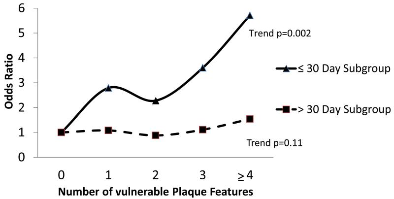
No significant heterogeneity between the studies was found. Predicted 5-year stroke risk (top versus bottom quartile) was related to plaque thrombus (odds ratio, 1.42; 95% confidence interval, 1.11-1.89; P=0.02), fibrous content (0.65; 0.49-0.87; P=0.004), macrophage infiltration (1.41; 1.05-1.90; P=0.02), high microvessel density (1.49; 1.05-2.11; P=0.03), and overall plaque instability (1.40; 1.05-1.87; P=0.02). This association was not observed for cap thickness, calcification, intraplaque hemorrhage, or lymphocyte infiltration. Plaques removed within 30 days of most recent symptomatic event were most strongly correlated with predicted stroke risk.
Features of the vulnerable carotid plaque, including plaque thrombus, low fibrous content, macrophage infiltration, and microvessel density, correlate with predicted stroke risk. This study provides a basis for plaque imaging studies focused on stroke risk stratification.
对于有症状的颈动脉狭窄患者,手术干预的风险效益在不同患者群体中可能有所不同。各种斑块成像方式已被推广为进行额外风险分层的潜在工具,尤其是在中度狭窄患者中。然而,颈动脉斑块成分在多大程度上可预测未来同侧缺血性卒中的风险仍不确定。
在两项大型动脉粥样硬化性颈动脉斑块生物样本库研究中,我们将1640个颈动脉斑块的组织学特征与一个经过验证的风险模型相关联,以预测个体1年和5年的卒中风险。
未发现两项研究之间存在显著异质性。预测的5年卒中风险(四分位数最高组与最低组相比)与斑块内血栓有关(比值比,1.42;95%置信区间,1.11 - 1.89;P = 0.02)、纤维成分(0.65;0.49 - 0.87;P = 0.004)、巨噬细胞浸润(1.41;1.05 - 1.90;P = 0.02)、微血管高密度(1.49;1.05 - 2.11;P = 0.03)以及总体斑块不稳定性(1.40;1.05 - 1.87;P = 0.02)。在帽厚度、钙化、斑块内出血或淋巴细胞浸润方面未观察到这种关联。在最近一次有症状事件发生后30天内切除的斑块与预测的卒中风险相关性最强。
易损颈动脉斑块的特征,包括斑块内血栓、低纤维成分、巨噬细胞浸润和微血管密度,与预测的卒中风险相关。本研究为专注于卒中风险分层的斑块成像研究提供了基础。