Tawakoli Pune N, Neu Thomas R, Busck Mette M, Kuhlicke Ute, Schramm Andreas, Attin Thomas, Wiedemeier Daniel B, Schlafer Sebastian
Clinic of Preventive Dentistry, Periodontology and Cardiology, Centre of Dental Medicine, University of Zurich, Zurich, Switzerland.
Department of River Ecology, Helmholtz Centre for Environmental Research - UFZ, Magdeburg, Germany.
J Oral Microbiol. 2017 Jul 9;9(1):1345581. doi: 10.1080/20002297.2017.1345581. eCollection 2017.
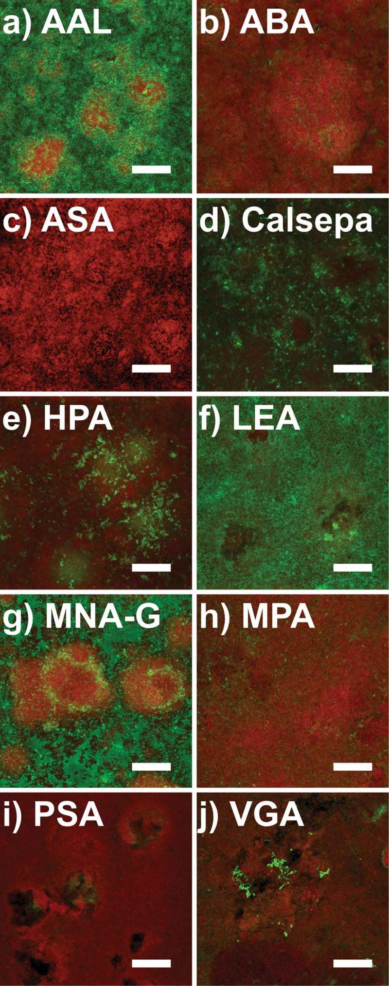
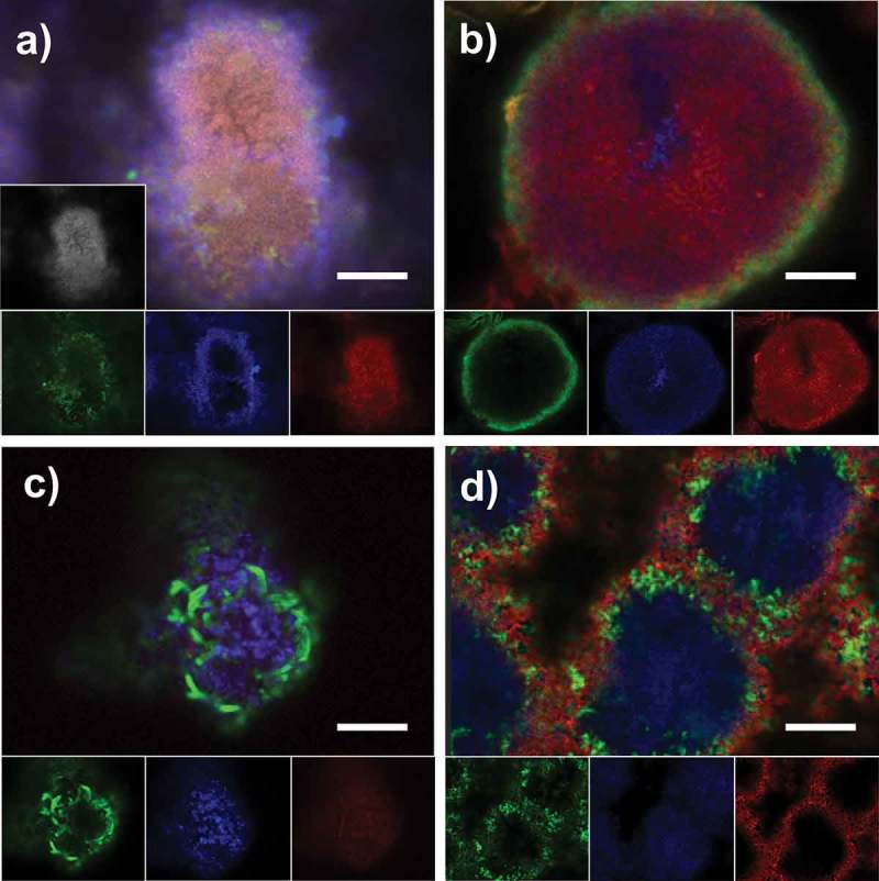
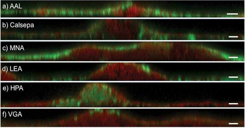
The extracellular matrix is a poorly studied, yet important component of dental biofilms. Fluorescence lectin-binding analysis (FLBA) is a powerful tool to characterize glycoconjugates in the biofilm matrix. This study aimed to systematically investigate the ability of 75 fluorescently labeled lectins to visualize and quantify extracellular glycoconjugates in dental biofilms. Lectin binding was screened on pooled supragingival biofilm samples collected from 76 subjects using confocal microscopy. FLBA was then performed with 10 selected lectins on biofilms grown for 48 h in the absence of sucrose. For five lectins that proved particularly suitable, stained biovolumes were quantified and correlated to the bacterial composition of the biofilms. Additionally, combinations of up to three differently labeled lectins were tested. Of the 10 lectins, five bound particularly well in 48-h-biofilms: (AAL), (Calsepa), (LEA), (MNA-G) and (HPA). No significant correlation between the binding of specific lectins and bacterial composition was found. Fluorescently labeled lectins enable the visualization of glycoconjugates in the dental biofilm matrix. The characterization and quantification of glycoconjugates in dental biofilms require a combination of several lectins. For 48-h-biofilms grown in absence of sucrose, AAL, Calsepa, HPA, LEA, and MNA-G are recommendable.
细胞外基质是牙菌斑中一个研究较少但很重要的成分。荧光凝集素结合分析(FLBA)是表征生物膜基质中糖缀合物的有力工具。本研究旨在系统研究75种荧光标记凝集素可视化和定量牙菌斑中细胞外糖缀合物的能力。使用共聚焦显微镜对从76名受试者收集的龈上生物膜混合样本进行凝集素结合筛选。然后用10种选定的凝集素对在无蔗糖条件下培养48小时的生物膜进行FLBA。对于五种被证明特别合适的凝集素,对染色的生物体积进行定量,并与生物膜的细菌组成相关联。此外,还测试了多达三种不同标记凝集素的组合。在这10种凝集素中,有五种在48小时生物膜中结合特别好:(AAL)、(Calsepa)、(LEA)、(MNA-G)和(HPA)。未发现特定凝集素的结合与细菌组成之间存在显著相关性。荧光标记凝集素能够可视化牙菌斑生物膜基质中的糖缀合物。牙菌斑中糖缀合物的表征和定量需要几种凝集素的组合。对于在无蔗糖条件下培养48小时的生物膜,推荐使用AAL、Calsepa、HPA、LEA和MNA-G。