• 文献检索
  • 文档翻译
  • 深度研究
  • 学术资讯
  • Suppr Zotero 插件Zotero 插件
  • 邀请有礼
  • 套餐&价格
  • 历史记录
应用&插件
Suppr Zotero 插件Zotero 插件浏览器插件Mac 客户端Windows 客户端微信小程序
定价
高级版会员购买积分包购买API积分包
服务
文献检索文档翻译深度研究API 文档MCP 服务
关于我们
关于 Suppr公司介绍联系我们用户协议隐私条款
关注我们

Suppr 超能文献

核心技术专利:CN118964589B侵权必究
粤ICP备2023148730 号-1Suppr @ 2026

文献检索

告别复杂PubMed语法,用中文像聊天一样搜索,搜遍4000万医学文献。AI智能推荐,让科研检索更轻松。

立即免费搜索

文件翻译

保留排版,准确专业,支持PDF/Word/PPT等文件格式,支持 12+语言互译。

免费翻译文档

深度研究

AI帮你快速写综述,25分钟生成高质量综述,智能提取关键信息,辅助科研写作。

立即免费体验

比较微计算机断层扫描成像与组织学在邻面龋检测中的应用。

Comparison of micro-CT imaging and histology for approximal caries detection.

机构信息

Department of Electronics and Informatics - ETRO Vrije Universiteit Brussel, Brussels, Belgium.

EXIA, Brussels, Belgium.

出版信息

Sci Rep. 2017 Jul 27;7(1):6680. doi: 10.1038/s41598-017-06735-6.

DOI:10.1038/s41598-017-06735-6
PMID:28751671
原文链接:https://pmc.ncbi.nlm.nih.gov/articles/PMC5532299/
Abstract

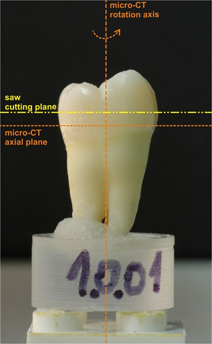
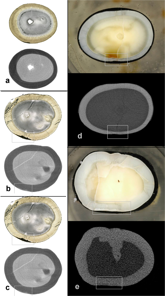
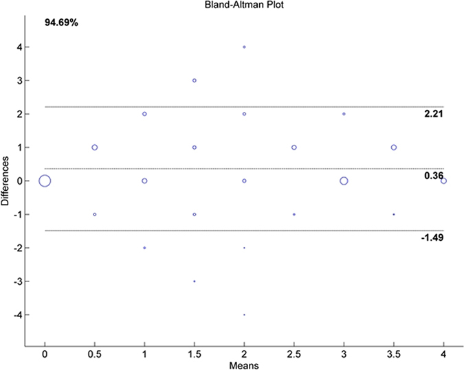
Histological sectioning is a generally accepted in vitro validation method for caries detection techniques. However, it requires cumbersome sample preparation and induces irreversible sample destruction. Micro-Computer Tomography (micro-CT) allows non-destructive imaging of tooth structure. The aim of this study was to compare the performance of histological sectioning and micro-CT imaging in detecting approximal carious lesions. Unlike previous studies, evaluation is objectified by comparing visual appearance of exactly corresponding anatomical regions. Sixty extracted human teeth were scanned with a desktop micro CT system. Axial histological slices were prepared and photographed. Sample preparation, combined with dedicated image processing, ensured selection of identical anatomical regions on radiographic and histological images. Evaluation of the presence and extent of carious lesions was performed by four dentists using custom-designed software. Each section was scored independently (histo or micro CT). Scores of approximal surfaces were retained for further analysis. Spearman's correlation coefficients (0.738 to 0.829, p < 0.0001) showed a good agreement between signs of carious lesions in the identical region obtained with both methods. Bland-Altman plots showed that 90.76% of the data points were within the limits of agreement. Micro-CT imaging was shown to provide an interesting alternative to histological sectioning as detection method for carious lesions.

摘要

组织学切片是一种被广泛接受的龋病检测技术的体外验证方法。然而,它需要繁琐的样本制备,并导致不可逆转的样本破坏。微计算机断层扫描(micro-CT)允许对牙齿结构进行非破坏性成像。本研究的目的是比较组织学切片和 micro-CT 成像在检测邻面龋损中的性能。与以往的研究不同,通过比较完全对应的解剖区域的视觉外观,使评估客观化。从 60 颗离体人牙中用台式 micro-CT 系统进行扫描。制备并拍摄轴向组织学切片。样本制备与专用图像处理相结合,确保在放射学和组织学图像上选择相同的解剖区域。四名牙医使用定制设计的软件评估龋损的存在和程度。每个切片分别进行评分(组织学或 micro-CT)。保留近中面的评分进行进一步分析。Spearman 相关系数(0.738 至 0.829,p < 0.0001)表明,两种方法在同一区域获得的龋损征象之间具有良好的一致性。Bland-Altman 图显示,90.76%的数据点在一致性界限内。micro-CT 成像被证明是一种有前途的替代组织学切片的方法,可作为龋病检测方法。

https://cdn.ncbi.nlm.nih.gov/pmc/blobs/1876/5532299/62ba20701436/41598_2017_6735_Fig3_HTML.jpg
https://cdn.ncbi.nlm.nih.gov/pmc/blobs/1876/5532299/94ac872f053b/41598_2017_6735_Fig1_HTML.jpg
https://cdn.ncbi.nlm.nih.gov/pmc/blobs/1876/5532299/e4f2cfad96bb/41598_2017_6735_Fig2_HTML.jpg
https://cdn.ncbi.nlm.nih.gov/pmc/blobs/1876/5532299/62ba20701436/41598_2017_6735_Fig3_HTML.jpg
https://cdn.ncbi.nlm.nih.gov/pmc/blobs/1876/5532299/94ac872f053b/41598_2017_6735_Fig1_HTML.jpg
https://cdn.ncbi.nlm.nih.gov/pmc/blobs/1876/5532299/e4f2cfad96bb/41598_2017_6735_Fig2_HTML.jpg
https://cdn.ncbi.nlm.nih.gov/pmc/blobs/1876/5532299/62ba20701436/41598_2017_6735_Fig3_HTML.jpg

相似文献

1
Comparison of micro-CT imaging and histology for approximal caries detection.比较微计算机断层扫描成像与组织学在邻面龋检测中的应用。
Sci Rep. 2017 Jul 27;7(1):6680. doi: 10.1038/s41598-017-06735-6.
2
In vitro comparison of D- and E-speed film radiography, RVG, and visualix digital radiography for the detection of enamel approximal and dentinal occlusal caries lesions.D速和E速胶片X线摄影、RVG及Visualix数字X线摄影用于检测釉质邻面和牙本质咬合面龋损的体外比较
Caries Res. 1994;28(5):363-7. doi: 10.1159/000262002.
3
The efficiency of three different films and radiovisiography in detecting approximal carious lesions.三种不同胶片及放射影像学在检测邻面龋损中的效能
Quintessence Int. 2005 Jan;36(1):65-70.
4
In vitro performance of near infrared light transillumination at 780-nm and digital radiography for detection of non-cavitated approximal caries.780nm 近红外光透射和数字射线照相术检测非窝沟性邻面龋的体外性能。
J Dent. 2017 Aug;63:44-50. doi: 10.1016/j.jdent.2017.05.018. Epub 2017 May 27.
5
Approximal caries depth assessment with storage phosphor versus film radiography. Evaluation of the caries-specific Oslo enhancement procedure.使用存储磷光体与胶片射线照相术进行邻面龋深度评估。龋病特异性奥斯陆增强程序的评价。
Caries Res. 2000 Nov-Dec;34(6):448-53. doi: 10.1159/000016622.
6
Comparison of diagnostic performance on approximal caries detection among Swedish and Chinese senior dental students using analogue and digital radiographs.瑞典和中国高年级牙科学生使用传统胶片和数字X线片检测邻面龋的诊断性能比较。
Swed Dent J. 2013;37(2):79-85.
7
Validity of MicroCT for in vitro detection of proximal carious lesions in primary molars.MicroCT 体外检测乳磨牙近中龋损的有效性。
J Dent. 2012 Jan;40(1):35-40. doi: 10.1016/j.jdent.2011.09.002. Epub 2011 Sep 12.
8
Influence of displayed image size on radiographic detection of approximal caries.显示图像大小对邻面龋X线片检测的影响。
Dentomaxillofac Radiol. 2003 Jul;32(4):242-6. doi: 10.1259/dmfr/17654484.
9
In vitro assessment of incipient approximal carious lesions using computer-assisted densitometric image analysis.使用计算机辅助密度测量图像分析对早期邻面龋损进行体外评估。
J Dent. 2002 Sep-Nov;30(7-8):305-11. doi: 10.1016/s0300-5712(02)00043-x.
10
In vitro comparison of conventional film and direct digital imaging in the detection of approximal caries.传统胶片与直接数字成像在邻面龋检测中的体外比较
Dentomaxillofac Radiol. 2007 Mar;36(3):138-42. doi: 10.1259/dmfr/15558921.

引用本文的文献

1
Enhancement of early proximal caries annotations in radiographs: introducing the Diagnostic Insights for Radiographic Early-caries with micro-CT (ACTA-DIRECT) dataset.增强放射影像中早期近中龋的标注:介绍基于微计算机断层扫描的放射影像早期龋诊断洞察(ACTA-DIRECT)数据集。
BMC Oral Health. 2024 Oct 30;24(1):1325. doi: 10.1186/s12903-024-05076-x.
2
Assessment of the Effect on Periodontitis of Antibiotic Therapy and Bacterial Lysate Treatment.评价抗生素治疗和细菌裂解物治疗对牙周炎的影响。
Int J Mol Sci. 2024 May 16;25(10):5432. doi: 10.3390/ijms25105432.
3
The effect of spatial resolution on deep learning classification of lung cancer histopathology.

本文引用的文献

1
Comparison of occlusal caries detection using the ICDAS criteria on extracted teeth or their photographs.使用国际龋病检测与评估系统(ICDAS)标准对拔除牙齿或其照片进行咬合面龋检测的比较。
BMC Oral Health. 2016 Sep 7;16(1):93. doi: 10.1186/s12903-016-0291-z.
2
Validation of micro-computed tomography for occlusal caries detection: an in vitro study.用于咬合面龋检测的微型计算机断层扫描技术的验证:一项体外研究。
Braz Oral Res. 2015;29(1):S1806-83242015000100309. doi: 10.1590/1807-3107BOR-2015.vol29.0132. Epub 2015 Oct 9.
3
Enamel lesion parameter correlations between polychromatic micro-CT and TMR.
空间分辨率对肺癌组织病理学深度学习分类的影响。
BJR Open. 2023 Aug 15;5(1):20230008. doi: 10.1259/bjro.20230008. eCollection 2023.
4
Proximal Caries Detection Using Short-Wave Infrared Transillumination at Wavelengths of 1050, 1200 and 1300 nm in Permanent Posterior Human Teeth.利用1050、1200和1300纳米波长的短波红外透照技术检测恒牙后牙的邻面龋
Diagnostics (Basel). 2023 Oct 19;13(20):3257. doi: 10.3390/diagnostics13203257.
5
Clinical Performance of Diagnostic Methods in Third Molar Teeth with Early Occlusal Caries.早期咬合龋第三磨牙诊断方法的临床性能
Diagnostics (Basel). 2023 Jan 12;13(2):284. doi: 10.3390/diagnostics13020284.
6
New Caries Diagnostic Tools in Intraoral Scanners: A Comparative In Vitro Study to Established Methods in Permanent and Primary Teeth.口腔内扫描仪中新龋病诊断工具的研究:与恒牙和乳牙中已建立方法的比较性体外研究。
Sensors (Basel). 2022 Mar 10;22(6):2156. doi: 10.3390/s22062156.
7
Micro-computed tomography in preventive and restorative dental research: A review.微计算机断层扫描在口腔预防与修复研究中的应用综述
Imaging Sci Dent. 2021 Dec;51(4):341-350. doi: 10.5624/isd.20210087. Epub 2021 Oct 15.
8
Near-infrared reflection at 780 nm for detection of early proximal caries in posterior permanent teeth in vitro.780nm 近红外反射用于体外探测后牙近中龋的早期病变。
Dentomaxillofac Radiol. 2021 Sep 1;50(6):20210005. doi: 10.1259/dmfr.20210005. Epub 2021 May 6.
9
Micro-computed tomography utility for estimation of intraparenchymal spinal cord cystic lesions in small animals.微计算机断层扫描在评估小动物实质内脊髓囊性病变中的应用
Neural Regen Res. 2021 Nov;16(11):2293-2298. doi: 10.4103/1673-5374.310690.
10
In vitro comparison of two types of digital X-ray sensors for proximal caries detection validated by micro-computed tomography.两种数字 X 射线传感器用于近端龋齿检测的体外比较,经微计算机断层扫描验证。
Dentomaxillofac Radiol. 2021 Mar 1;50(3):20200338. doi: 10.1259/dmfr.20200338. Epub 2020 Aug 27.
多色微 CT 与 TMR 之间的釉质病变参数相关性。
J Dent Res. 2012 Jun;91(6):586-91. doi: 10.1177/0022034512444127. Epub 2012 Apr 3.
4
Validity of MicroCT for in vitro detection of proximal carious lesions in primary molars.MicroCT 体外检测乳磨牙近中龋损的有效性。
J Dent. 2012 Jan;40(1):35-40. doi: 10.1016/j.jdent.2011.09.002. Epub 2011 Sep 12.
5
Artefacts in CBCT: a review.CBCT 中的伪影:综述。
Dentomaxillofac Radiol. 2011 Jul;40(5):265-73. doi: 10.1259/dmfr/30642039.
6
Does DIAGNOdent provide a reliable caries-removal endpoint?DIAGNOdent 能否提供可靠的龋齿去除终点?
J Dent. 2011 May;39(5):351-60. doi: 10.1016/j.jdent.2011.02.005. Epub 2011 Feb 18.
7
Application of scanning microradiography and x-ray microtomography to studies of bones and teeth.扫描显微射线照相术和 X 射线微断层摄影术在骨骼和牙齿研究中的应用。
J Xray Sci Technol. 1994 Jan 1;4(2):102-17. doi: 10.3233/XST-1994-4204.
8
An automated assessment algorithm for micro-CT images of occlusal caries.一种用于评估咬合面龋的 micro-CT 图像的自动化算法。
Surgeon. 2010 Dec;8(6):334-40. doi: 10.1016/j.surge.2010.06.007. Epub 2010 Jul 22.
9
Diagnostic performance of the visual caries classification system ICDAS II versus radiography and micro-computed tomography for proximal caries detection: an in vitro study.视觉龋分类系统 ICDAS II 与放射摄影和微计算机断层扫描在检测邻面龋中的诊断性能:一项体外研究。
J Dent. 2010 Nov;38(11):859-67. doi: 10.1016/j.jdent.2010.07.005. Epub 2010 Jul 21.
10
A system of calibrating microtomography for use in caries research.一种用于龋齿研究的显微断层扫描校准系统。
Caries Res. 2009;43(4):314-21. doi: 10.1159/000226230. Epub 2009 Jun 24.