Gao Yuan, Li Su, Xu Dazhi, Chen Shangxiang, Cai Yuchen, Jiang Wenqi, Zhang Xinke, Sun Jin, Wang Kefeng, Chang Boyang, Wang Fenghua, Hong Minghuang
Sun Yat-sen University Cancer Center; State Key Laboratory of Oncology in South China; Collaborative Innovation Center for Cancer Medicine, Guangzhou, 510060, Guangdong, P. R. China.
Department of Clinical Trial Center, Sun Yat-sen University Cancer Center, Guangzhou, 510060, Guangdong, P. R. China.
Chin J Cancer. 2017 Jul 29;36(1):61. doi: 10.1186/s40880-017-0226-3.
Anti-programmed death-1/programmed death-ligand 1 (PD-1/PD-L1) immunotherapy has been proved to be effective on gastric cancer in ongoing clinical trials. However, the value of PD-L1 in predicting responses of patients with gastric cancer to anti-PD-1/PD-L1 immunotherapy is controversial. Some studies suggested that intra- and inter-tumoral heterogeneity of PD-L1 expression might explain the controversy. This study aimed to analyze the expression of PD-L1, PD-L2, and PD-1 as well as CD8(+) T-cell density in primary tumors and lymph nodes from patients with stage T1-4N+M0 gastric adenocarcinoma to explore the heterogeneity of PD-1 signaling pathway molecules.
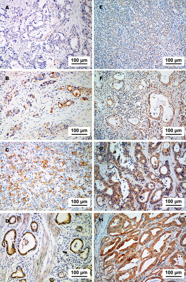
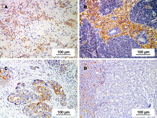
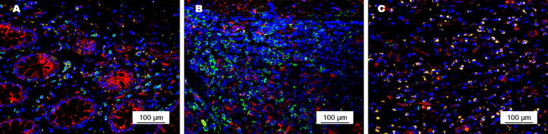
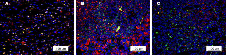
In primary tumors and metastatic as well as non-metastatic lymph nodes from patients with stage T1-4N+M0 gastric adenocarcinoma, we detected PD-L1 and PD-L2 expression with immunohistochemistry. CD8(+) T-cell density in primary tumors and PD-1 expression on CD8(+) T cells were detected with immunofluorescence. Univariate analysis was used to determine the prognostic values of them. Cox proportional hazard regression model was used to identify independent risk factors that affect patients' overall survival and disease-free survival.
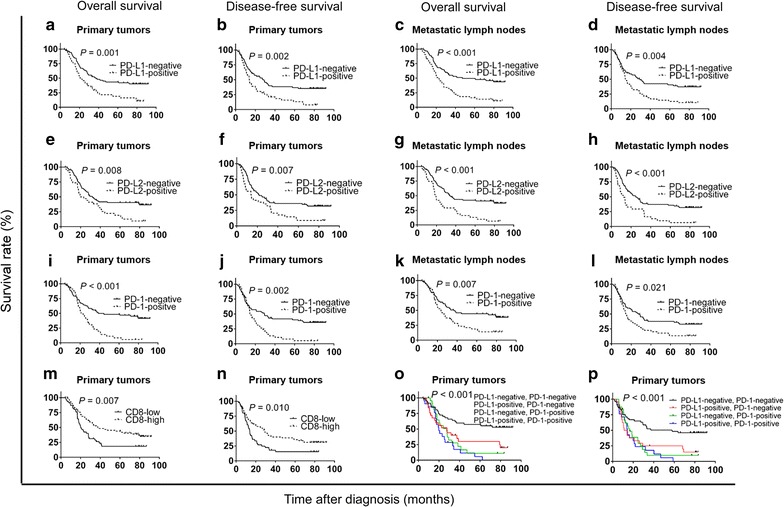
Among 119 eligible patients who had undergone surgical resection, the positive rate of PD-L1 was higher in metastatic lymph nodes than in primary tumors (45.4% vs. 38.7%, P = 0.005); the positive rate of PD-1 on CD8(+) T cells was significantly higher in primary tumors and metastatic lymph nodes than in tumor-free lymph nodes (both P < 0.001). The intensity of PD-1 expression on CD8(+) T cells in primary tumors and in metastatic lymph nodes were stronger than that in tumor-free lymph nodes from the same patient. Beside, the positive rate of PD-L2 did not show any differences between primary tumors and metastatic lymph nodes. In multivariate analysis, PD-L1 expression, PD-L2 expression, a low density of CD8(+) T cells in primary tumors, and PD-1 expression on CD8(+) T cells in primary tumors were associated with poor prognosis.
The expression of PD-L1 is heterogeneous in primary tumors and in metastatic lymph nodes from patients with stage T1-4N+M0 gastric adenocarcinoma, which might explain the inconsistent results in assessing the prognostic value of PD-L1 expression in previous studies.
在正在进行的临床试验中,抗程序性死亡蛋白1/程序性死亡配体1(PD-1/PD-L1)免疫疗法已被证明对胃癌有效。然而,PD-L1在预测胃癌患者对抗PD-1/PD-L1免疫疗法反应中的价值存在争议。一些研究表明,PD-L1表达的肿瘤内和肿瘤间异质性可能解释了这一争议。本研究旨在分析T1-4N+M0期胃腺癌患者原发肿瘤和淋巴结中PD-L1、PD-L2和PD-1的表达以及CD8(+) T细胞密度,以探讨PD-1信号通路分子的异质性。
在T1-4N+M0期胃腺癌患者的原发肿瘤、转移及非转移淋巴结中,我们采用免疫组化检测PD-L1和PD-L2表达。采用免疫荧光检测原发肿瘤中CD8(+) T细胞密度及CD8(+) T细胞上的PD-1表达。采用单因素分析确定它们的预后价值。采用Cox比例风险回归模型确定影响患者总生存和无病生存的独立危险因素。
在119例接受手术切除的合格患者中,转移淋巴结中PD-L1阳性率高于原发肿瘤(45.4%对38.7%,P = 0.005);原发肿瘤和转移淋巴结中CD8(+) T细胞上PD-1阳性率显著高于无瘤淋巴结(均P < 0.001)。同一患者原发肿瘤和转移淋巴结中CD8(+) T细胞上PD-1表达强度强于无瘤淋巴结。此外,原发肿瘤和转移淋巴结中PD-L2阳性率无差异。多因素分析显示,PD-L1表达、PD-L2表达、原发肿瘤中CD8(+) T细胞低密度以及原发肿瘤中CD8(+) T细胞上PD-1表达与预后不良相关。
T1-4N+M0期胃腺癌患者原发肿瘤和转移淋巴结中PD-L1表达存在异质性,这可能解释了既往研究中评估PD-L1表达预后价值结果不一致的原因。