Department of Urology, West China Hospital, Sichuan University, Chengdu, People's Republic of China, 610041.
Institute of Urology, West China Hospital, Sichuan University, Chengdu, People's Republic of China, 610041.
BMC Cancer. 2019 Apr 16;19(1):360. doi: 10.1186/s12885-019-5578-4.
In clinical practice, the detection of biomarkers is mostly based on primary tumors for its convenience in acquisition. However, immune checkpoints may express differently between primary and metastatic tumor. Therefore, we aimed to compare the differential expressions of PD-1, PD-L1 and PD-L2 between the primary and metastatic sites of renal cell carcinoma (RCC).
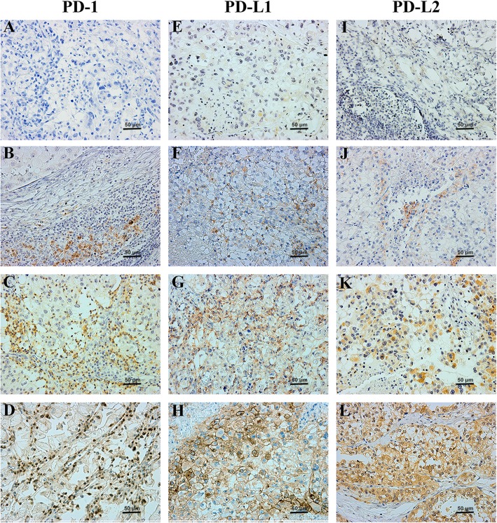
Patients diagnosed with RCC by resection or fine needle aspiration of metastasis were included. Immunohistochemistry (IHC) was applied to detect PD-1, PD-L1 and PD-L2 expressions. SPSS 22.0 was applied to conduct Chi-square, consistency tests and Cox's proportional hazards regression models. GraphPad Prism 6 was used to plot survival curves and R software was used to calculate Predictive accuracy (PA).
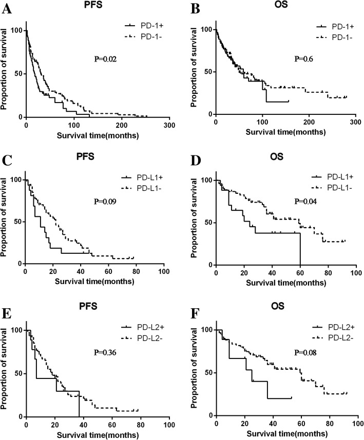
In the whole cohort (N = 163), IHC results suggested a higher detection rate of PD-L1 in the metastasis than that of the primary site (χ2 = 4.66, p = 0.03), with a low consistent rate of 32.5%. Among different metastatic tumors, PD-1 was highly expressed in the lung/lymph node (65.3%) and poorly expressed in the brain (10.5%) and visceral metastases (12.5%). PD-L1 was highly expressed in lung/lymph node (37.5%) and the bone metastases (12.2%) on the contrary. In terms of survival analysis, patients with PD-1 expression either in the primary or metastasis had a shorter overall survival (OS) (HR: 1.59, 95% CI 1.08-2.36, p = 0.02). Also, PD-L1 expression in the primary was associated with a shorter OS (HR 2.55, 95% CI 1.06-6.15, p = 0.04). In the multivariate analysis, the predictive accuracy of the whole model for PFS was increased from 0.683 to 0.699 after adding PD-1.
PD-1, PD-L1 and PD-L2 were differentially expressed between primary and metastatic tumors. Histopathological examination of these immune check points in metastatic lesions of mRCC should be noticed, and its accurate diagnosis may be one of the effective ways to realize the individualized treatment.
在临床实践中,生物标志物的检测大多基于原发肿瘤,因为其获取较为方便。然而,免疫检查点在原发肿瘤和转移肿瘤之间的表达可能不同。因此,我们旨在比较肾细胞癌(RCC)原发灶和转移灶之间 PD-1、PD-L1 和 PD-L2 的差异表达。
纳入通过转移灶切除术或细针抽吸术诊断为 RCC 的患者。应用免疫组织化学(IHC)检测 PD-1、PD-L1 和 PD-L2 的表达。采用 SPSS 22.0 进行卡方检验、一致性检验和 Cox 比例风险回归模型。采用 GraphPad Prism 6 绘制生存曲线,采用 R 软件计算预测准确率(PA)。
在整个队列(N=163)中,IHC 结果提示转移灶中 PD-L1 的检出率高于原发灶(χ2=4.66,p=0.03),一致性率较低,为 32.5%。在不同的转移肿瘤中,PD-1 在肺/淋巴结(65.3%)中高表达,在脑(10.5%)和内脏转移(12.5%)中低表达。PD-L1 在肺/淋巴结(37.5%)和骨转移(12.2%)中高表达。在生存分析方面,原发灶或转移灶中 PD-1 表达的患者总生存(OS)更短(HR:1.59,95%CI 1.08-2.36,p=0.02)。此外,原发灶 PD-L1 表达与 OS 更短相关(HR 2.55,95%CI 1.06-6.15,p=0.04)。在多变量分析中,在加入 PD-1 后,整个模型对 PFS 的预测准确率从 0.683 提高到 0.699。
PD-1、PD-L1 和 PD-L2 在原发灶和转移灶之间存在差异表达。在转移性 mRCC 的免疫检查点的组织病理学检查中应注意这些变化,其准确诊断可能是实现个体化治疗的有效途径之一。