Louisraj Sophia, Ponnudurai Thendral, Rodriguez Dominic, Thomas Philip A, Nelson Jesudasan Christadoss Arul
Department of Orbit and Oculoplasty, Joseph Eye Hospital.
Department of Medicine, Kauvery Medical Centre, Tiruchirapalli, India.
Int Med Case Rep J. 2017 Jul 24;10:255-259. doi: 10.2147/IMCRJ.S133284. eCollection 2017.
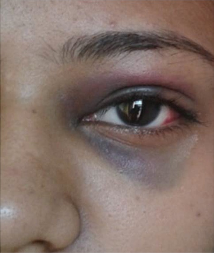
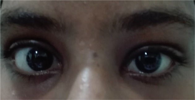
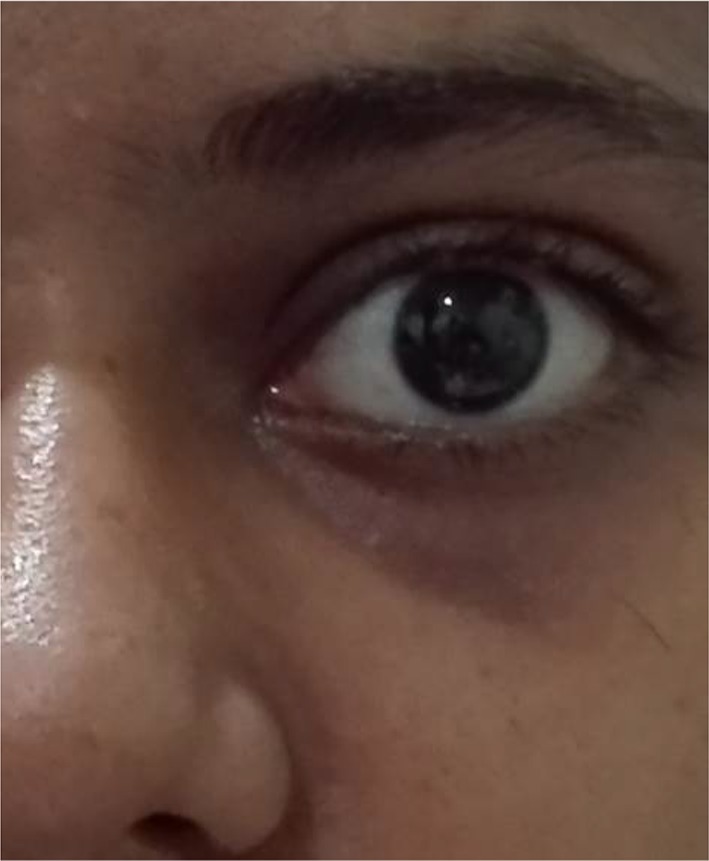
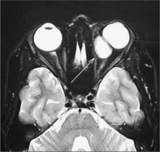
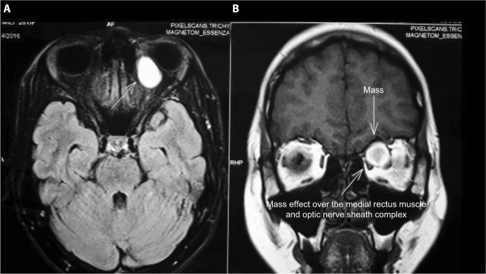
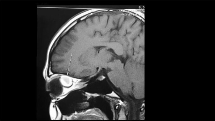
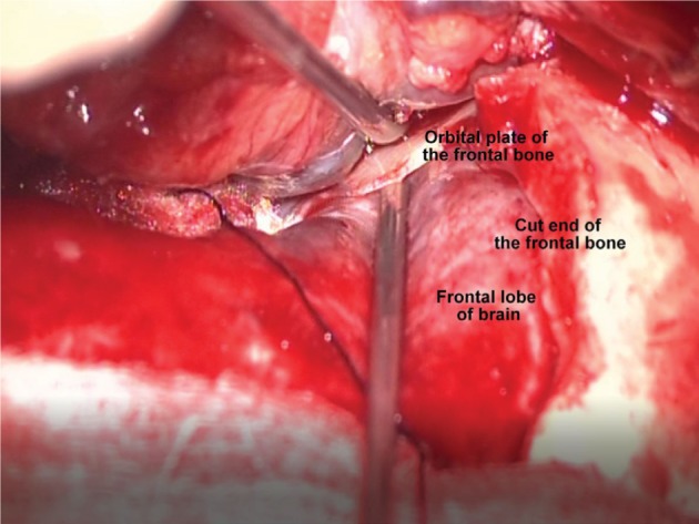
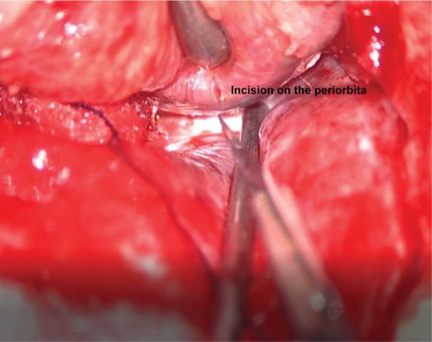
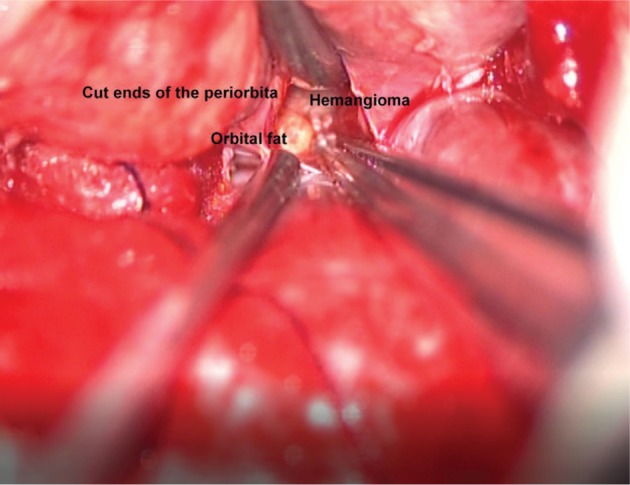
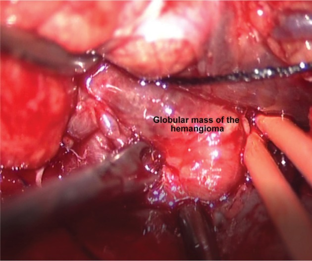
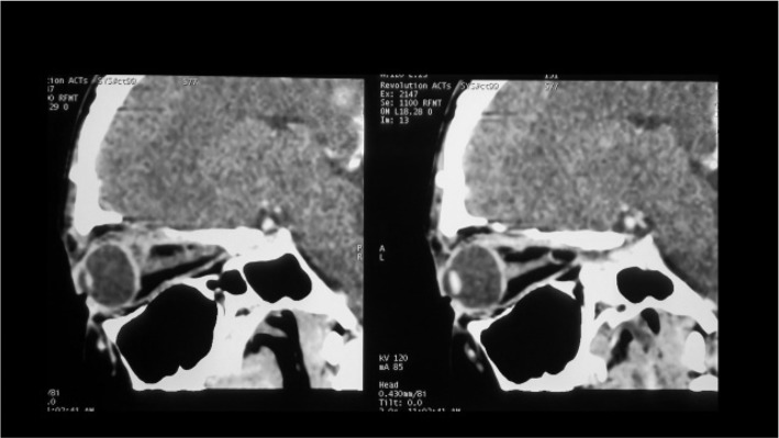
We report an unusual presentation of an orbital cavernous hemangioma in a 26-year-old female, who noted sudden redness and swelling of the left eye (LE) on waking up. At presentation, upper eyelid edema with periorbital ecchymosis and subconjunctival hemorrhage were noted in the LE. Although there was transient symptomatic relief with topical medications, blurring of vision developed in the LE. When seen 10 days later, the patient's LE showed axial proptosis. Magnetic resonance imaging revealed an intraconal soft tissue mass in the superomedial quadrant of the left orbit. Superior orbitotomy with mass excision was done; histopathological examination of the excised mass revealed a cavernous hemangioma. The patient had complete visual recovery following surgery. To our knowledge, an acute presentation of an orbital cavernous hemangioma with subconjunctival hemorrhage and periorbital ecchymosis has not previously been reported.
我们报告了一例26岁女性眼眶海绵状血管瘤的不寻常表现,该患者醒来时发现左眼突然发红肿胀。就诊时,左眼可见上睑水肿伴眶周瘀斑及结膜下出血。尽管局部用药后症状有短暂缓解,但左眼出现视力模糊。10天后复诊时,患者左眼出现轴性眼球突出。磁共振成像显示左眼眶上内侧象限锥内软组织肿块。行眶上切开术并切除肿块;切除肿块的组织病理学检查显示为海绵状血管瘤。患者术后视力完全恢复。据我们所知,眼眶海绵状血管瘤伴结膜下出血和眶周瘀斑的急性表现此前未见报道。