Martineau Xavier, Abed Élie, Martel-Pelletier Johanne, Pelletier Jean-Pierre, Lajeunesse Daniel
Unité de recherche en Arthrose, Centre de recherche du Centre hospitalier de l'Université de Montréal (CRCHUM), Montréal, Québec, Canada.
PLoS One. 2017 Aug 4;12(8):e0180711. doi: 10.1371/journal.pone.0180711. eCollection 2017.
Clinical and in vitro studies suggest that subchondral bone sclerosis due to abnormal osteoblasts (Ob) is involved in the progression and/or onset of osteoarthritis (OA). Human Ob isolated from sclerotic subchondral OA bone tissue show an altered phenotype, a decreased canonical Wnt/β-catenin signaling pathway (cWnt), and a reduced mineralization in vitro. In addition to the cWnt pathway, at least two non-canonical signaling pathways, the Wnt/PKC and Wnt/PCP pathway have been described. However, there are no reports of either pathway in OA Ob. Here, we studied the two non-canonical pathways in OA Ob and if they influence their phenotype.
Human primary subchondral Ob were isolated from the subchondral bone plate of tibial plateaus of OA patients undergoing total knee arthroplasty, or of normal individuals at autopsy. The expression of genes involved in non-canonical Wnt signaling was evaluated by qRT-PCR and their protein production by Western blot analysis. Alkaline phosphatase activity and osteocalcin secretion (OC) were determined with substrate hydrolysis and EIA, respectively. Mineralization levels were evaluated with Alizarin Red Staining, Wnt/PKC and Wnt/PCP pathways by target gene expression and their respective activity using the NFAT and AP-1 luciferase reporter assays.
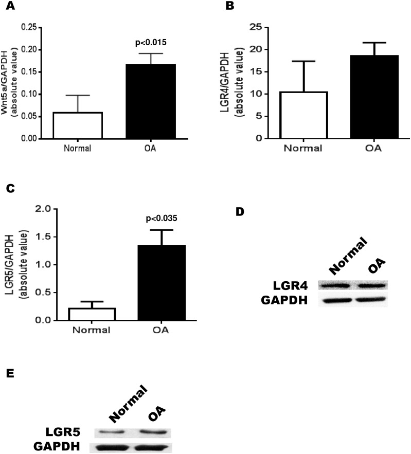
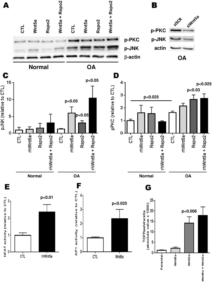
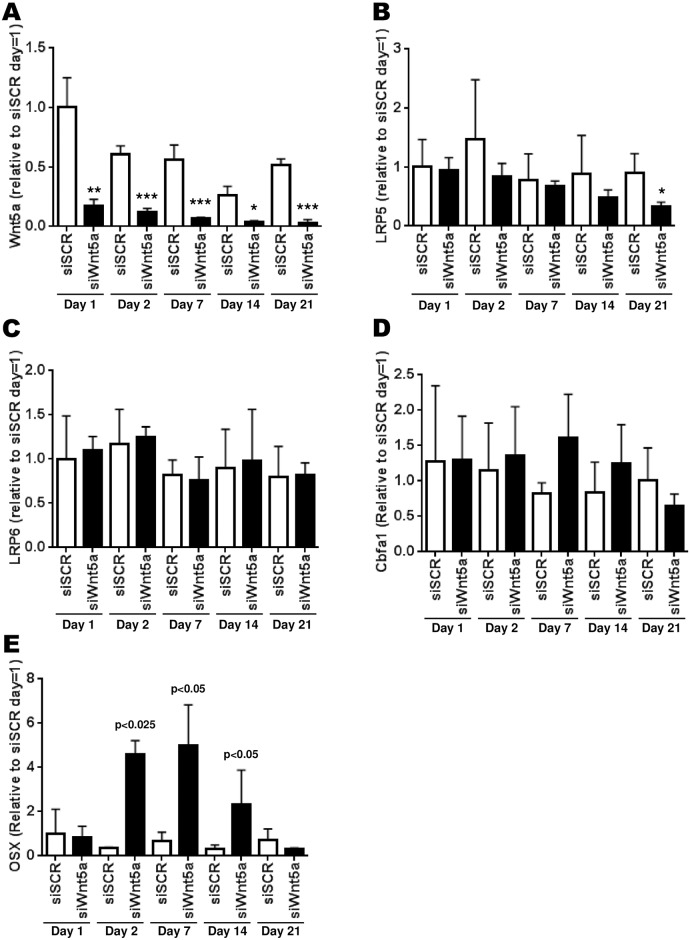
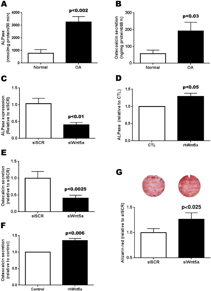
OA Ob showed an altered phenotype as illustrated by an increased alkaline phosphatase activity and osteocalcin release compared to normal Ob. The expression of the non-canonical Wnt5a ligand was increased in OA Ob compared to normal. Whereas, the expression of LGR5 was significantly increased in OA Ob compared to normal Ob, the expression of LGR4 was similar. Wnt5a directly stimulated the expression and production of LGR5, contrasting, Wnt5a did not stimulate the expression of LGR4. Wnt5a also stimulated the phosphorylation of both JNK and PKC, as well as the activity of both NFAT and AP-1 transcription factors. The inhibition of Wnt5a expression partially corrects the abnormal mineralization, OC secretion and ALPase activity of OA Ob.
These data indicate that the alteration of Wnt5a, a non-canonical Wnt signaling activator, is implicated in the modified signalisation and phenotype observed in OA Ob.
临床及体外研究表明,成骨细胞(Ob)异常导致的软骨下骨硬化参与了骨关节炎(OA)的进展和/或发病过程。从硬化的OA软骨下骨组织中分离出的人Ob表现出表型改变、经典Wnt/β-连环蛋白信号通路(cWnt)降低以及体外矿化减少。除了cWnt通路外,还描述了至少两种非经典信号通路,即Wnt/PKC和Wnt/PCP通路。然而,尚无关于OA Ob中这两种通路的报道。在此,我们研究了OA Ob中的两种非经典通路及其是否影响其表型。
从接受全膝关节置换术的OA患者或尸检正常个体的胫骨平台软骨下骨板中分离出人原发性软骨下Ob。通过qRT-PCR评估参与非经典Wnt信号通路的基因表达,并通过蛋白质印迹分析评估其蛋白质产生。分别用底物水解和酶免疫分析测定碱性磷酸酶活性和骨钙素分泌(OC)。用茜素红染色评估矿化水平,通过靶基因表达以及使用NFAT和AP-1荧光素酶报告基因测定法评估Wnt/PKC和Wnt/PCP通路及其各自的活性。
与正常Ob相比,OA Ob表现出表型改变,表现为碱性磷酸酶活性增加和骨钙素释放增加。与正常Ob相比,OA Ob中非经典Wnt5a配体的表达增加。然而,与正常Ob相比,OA Ob中LGR5的表达显著增加,而LGR4的表达相似。Wnt5a直接刺激LGR5的表达和产生,相反,Wnt5a不刺激LGR4的表达。Wnt5a还刺激JNK和PKC的磷酸化以及NFAT和AP-1转录因子的活性。抑制Wnt5a表达可部分纠正OA Ob的异常矿化、OC分泌和碱性磷酸酶活性。
这些数据表明,非经典Wnt信号激活剂Wnt5a的改变与OA Ob中观察到的信号转导改变和表型有关。