Unité de recherche en Arthose, Centre de recherche du Centre Hospitalier de l'Université de Montréal (CR-CHUM), Hôpital Notre-Dame, 1560 rue Sherbrooke Est, Montréal, QC H2L 4 M1, Canada.
Arthritis Res Ther. 2010;12(1):R20. doi: 10.1186/ar2925. Epub 2010 Feb 8.
Leptin is a peptide hormone with a role in bone metabolism and rheumatic diseases. The subchondral bone tissue plays a prominent role in the pathophysiology of osteoarthritis (OA), related to abnormal osteoblast (Ob) differentiation. Although leptin promotes the differentiation of Ob under normal conditions, a role for leptin in OA Ob has not been demonstrated. Here we determined if endogenous leptin produced by OA Ob could be responsible for the expression of the abnormal phenotypic biomarkers observed in OA Ob.
We prepared primary normal and OA Ob from subchondral bone of tibial plateaus removed for knee surgery of OA patients or at autopsy. We determined the production of leptin and of the long, biologically active, leptin receptors (OB-Rb) using reverse transcriptase-polymerase chain reaction, ELISA and Western blot analysis. We determined the effect of leptin on cell proliferation by BrdU incorporation and 3-(4,5-Dimethylthiazol-2-yl)-2,5-diphenyltetrazolium bromide (MTT) assays, and we determined by Western blot analysis phospho 42/44 MAPK (p42/44 Erk1/2) and phospho p38 levels. We then determined the effect of the addition of exogenous leptin, leptin receptor antagonists, inhibitors of leptin signaling or siRNA techniques on the phenotypic features of OA Ob. Phenotypic features of Ob were determined by measuring alkaline phosphatase activity (ALP), osteocalcin release (OC), collagen type 1 production (CICP) and of Transforming Growth Factor-beta1 (TGF-beta1).
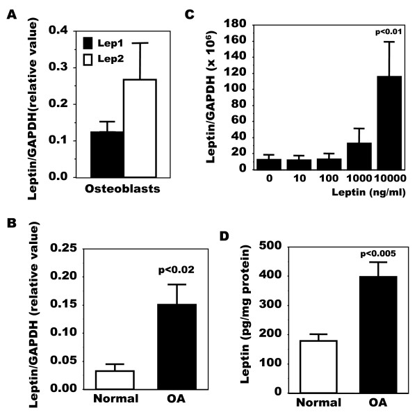
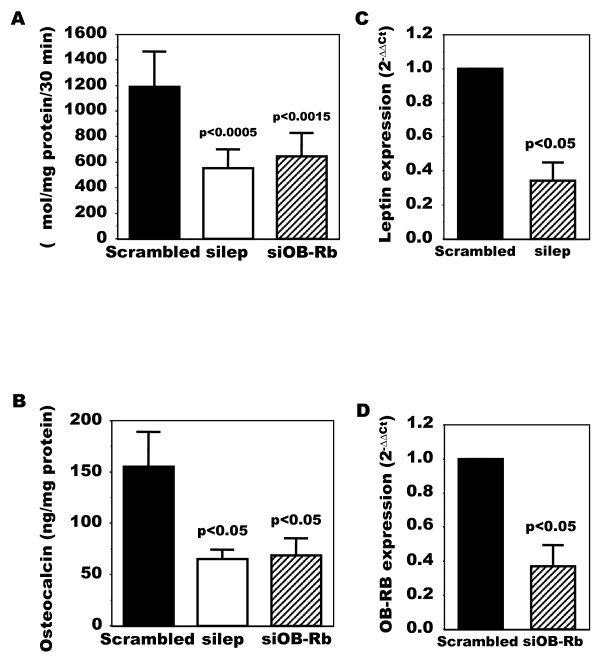
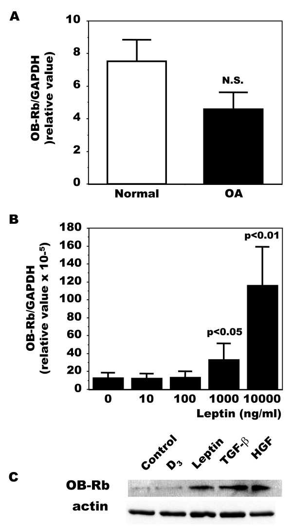
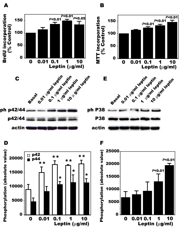
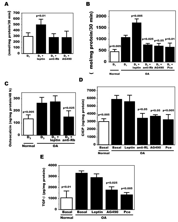
Leptin expression was increased approximately five-fold and protein levels approximately two-fold in OA Ob compared to normal. Leptin stimulated its own expression and the expression of OB-Rb in OA Ob. Leptin dose-dependently stimulated cell proliferation of OA Ob and also increased phosphorylated p42/44 Erk1/2 and p38 levels. Inactivating antibodies against leptin reduced ALP, OC, CICP and TGF-beta1 levels in OA Ob. Tyrphostin (AG490) and piceatannol (Pce), inhibitors of leptin signaling, reproduced this effect. Inhibition of endogenous leptin levels using siRNA for leptin or inhibiting leptin signaling using siRNA for OB-Rb expression both reduced ALP and OC about 60%. Exogenous leptin addition stimulated ALP, yet this failed to further increase OC or CICP.
These results suggest that abnormal production of leptin by OA Ob could be responsible, in part, for the elevated levels of ALP, OC, collagen type 1 and TGF-beta1 observed in these cells compared to normal. Leptin also stimulated cell proliferation, and Erk 1/2 and p38 signaling. Taken together, these data suggest leptin could contribute to abnormal osteoblast function in OA.
瘦素是一种在骨骼代谢和风湿性疾病中起作用的肽激素。软骨下骨组织在骨关节炎(OA)的病理生理学中起着突出的作用,与成骨细胞(Ob)分化异常有关。尽管瘦素在正常情况下促进 Ob 的分化,但瘦素在 OA Ob 中的作用尚未得到证实。在这里,我们确定 OA Ob 产生的内源性瘦素是否可能是 OA Ob 中观察到的异常表型生物标志物表达的原因。
我们从 OA 患者膝关节手术或尸检切除的胫骨平台软骨下骨中制备原代正常和 OA Ob。我们使用逆转录酶聚合酶链反应、ELISA 和 Western blot 分析来确定瘦素和长的、具有生物活性的瘦素受体(OB-Rb)的产生。我们通过 BrdU 掺入和 3-(4,5-二甲基噻唑-2-基)-2,5-二苯基四唑溴盐(MTT)测定来确定瘦素对细胞增殖的影响,并用 Western blot 分析测定磷酸 42/44 MAPK(p42/44 Erk1/2)和磷酸化 p38 水平。然后,我们确定添加外源性瘦素、瘦素受体拮抗剂、瘦素信号抑制剂或 siRNA 技术对 OA Ob 表型特征的影响。Ob 的表型特征通过测量碱性磷酸酶活性(ALP)、骨钙素释放(OC)、胶原蛋白 1 产生(CICP)和转化生长因子-β1(TGF-β1)来确定。
与正常 Ob 相比,OA Ob 中的瘦素表达增加约五倍,蛋白水平增加约两倍。瘦素刺激 OA Ob 自身及其 OB-Rb 的表达。瘦素以剂量依赖的方式刺激 OA Ob 的细胞增殖,并且还增加了磷酸化的 p42/44 Erk1/2 和 p38 水平。针对瘦素的中和抗体减少了 OA Ob 中的 ALP、OC、CICP 和 TGF-β1 水平。瘦素信号的抑制剂 Tyrphostin(AG490)和 Piceatannol(Pce)复制了这种效果。使用瘦素 siRNA 或 OB-Rb 表达的 siRNA 抑制内源性瘦素水平均使 ALP 和 OC 降低约 60%。外源性瘦素的添加刺激了 ALP,但未能进一步增加 OC 或 CICP。
这些结果表明,OA Ob 异常产生的瘦素可能部分导致与正常相比,这些细胞中观察到的 ALP、OC、胶原蛋白 1 和 TGF-β1 水平升高。瘦素还刺激细胞增殖和 Erk 1/2 和 p38 信号。综上所述,这些数据表明瘦素可能有助于 OA 中异常成骨细胞功能。