Amarnani Dhanesh, Machuca-Parra Arturo Israel, Wong Lindsay L, Marko Christina K, Stefater James A, Stryjewski Tomasz P, Eliott Dean, Arboleda-Velasquez Joseph F, Kim Leo A
Schepens Eye Research Institute of Massachusetts Eye and Ear, Boston, Massachusetts, United States.
Retina Service, Massachusetts Eye and Ear, Department of Ophthalmology, Harvard Medical School, Boston, Massachusetts, United States.
Invest Ophthalmol Vis Sci. 2017 Aug 1;58(10):3940-3949. doi: 10.1167/iovs.16-20912.
The purpose of this study was to develop a method for isolating, culturing, and characterizing cells from patient-derived membranes in proliferative vitreoretinopathy (PVR) to be used for drug testing.
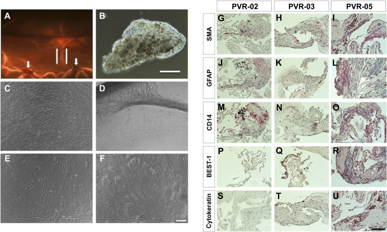
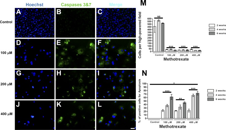
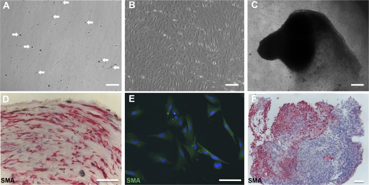
PVR membranes were obtained from six patients with grade C PVR. Membrane fragments were analyzed by gross evaluation, fixed for immunohistologic studies to establish cell identity, or digested with collagenase II to obtain single cell suspensions for culture. PVR-derived primary cultures were used to examine the effects of methotrexate (MTX) on proliferation, migration, and cell death.
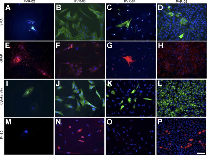
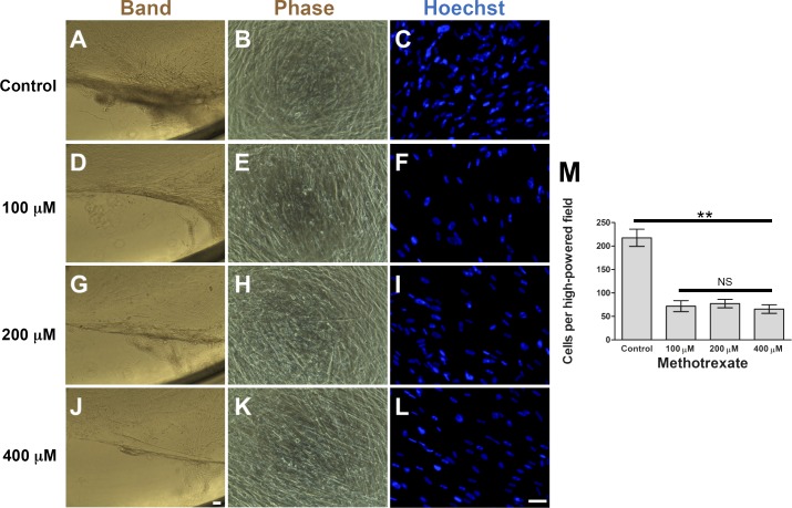
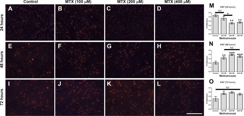
Gross analysis of PVR membranes showed presence of pigmented cells, indicative of retinal pigment epithelial cells. Immunohistochemistry identified cells expressing α-smooth muscle actin, glial fibrillary acidic protein, Bestrophin-1, and F4/80, suggesting the presence of multiple cell types in PVR. Robust PVR primary cultures (C-PVR) were successfully obtained from human membranes, and these cells retained the expression of cell identity markers in culture. C-PVR cultures formed membranes and band-like structures in culture reminiscent of the human condition. MTX significantly reduced the proliferation and band formation of C-PVR, whereas it had no significant effect on cell migration. MTX also induced regulated cell death within C-PVR as assessed by increased expression of caspase-3/7.
PVR cells obtained from human membranes can be successfully isolated, cultured, and profiled in vitro. Using these primary cultures, we identified MTX as capable of significantly reducing growth and inducing cell death of PVR cells in vitro.
本研究的目的是开发一种从增殖性玻璃体视网膜病变(PVR)患者来源的膜中分离、培养和鉴定细胞的方法,用于药物测试。
从6例C级PVR患者获取PVR膜。通过大体评估分析膜碎片,固定用于免疫组织学研究以确定细胞身份,或用胶原酶II消化以获得单细胞悬液用于培养。使用PVR来源的原代培养物检测甲氨蝶呤(MTX)对增殖、迁移和细胞死亡的影响。
PVR膜的大体分析显示存在色素细胞,提示视网膜色素上皮细胞。免疫组织化学鉴定出表达α-平滑肌肌动蛋白、胶质纤维酸性蛋白、Bestrophin-1和F4/80的细胞,表明PVR中存在多种细胞类型。成功从人膜中获得了强大的PVR原代培养物(C-PVR),并且这些细胞在培养中保留了细胞身份标志物的表达。C-PVR培养物在培养中形成了类似于人类情况的膜和带状结构。MTX显著降低了C-PVR的增殖和带状结构形成,而对细胞迁移没有显著影响。通过增加的caspase-3/7表达评估,MTX还诱导了C-PVR内的程序性细胞死亡。
从人膜中获得的PVR细胞可以在体外成功分离、培养和分析。使用这些原代培养物,我们确定MTX能够在体外显著降低PVR细胞的生长并诱导细胞死亡。