Dvashi Zeev, Goldberg Mordechai, Adir Orit, Shapira Michal, Pollack Ayala
Kaplan Medical Center, Rehovot, affiliated with Hadassah-Hebrew University of Jerusalem, Rehovot, Israel.
PLoS One. 2015 Apr 7;10(4):e0122229. doi: 10.1371/journal.pone.0122229. eCollection 2015.
Proliferative vitreoretinopathy (PVR) is an active process that develops as a complication upon retinal detachment (RD), accompanied by formation of fibrotic tissue. The main cells involved in the development of fibrotic tissue during PVR are the retinal pigment epithelial (RPE) cells. The RPE cells undergo epithelial-mesenchymal transition (EMT) which leads to complex retinal detachment and loss of vision. Transforming growth factor-β1 (TGF-β1) is considered as the main player in the EMT of RPE cells, even though the mechanism is not fully understood. This study was performed to determine the possible involvement of transforming growth factor β activated kinase 1 (TAK1) in the EMT process of the RPE cells.
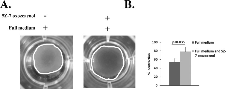
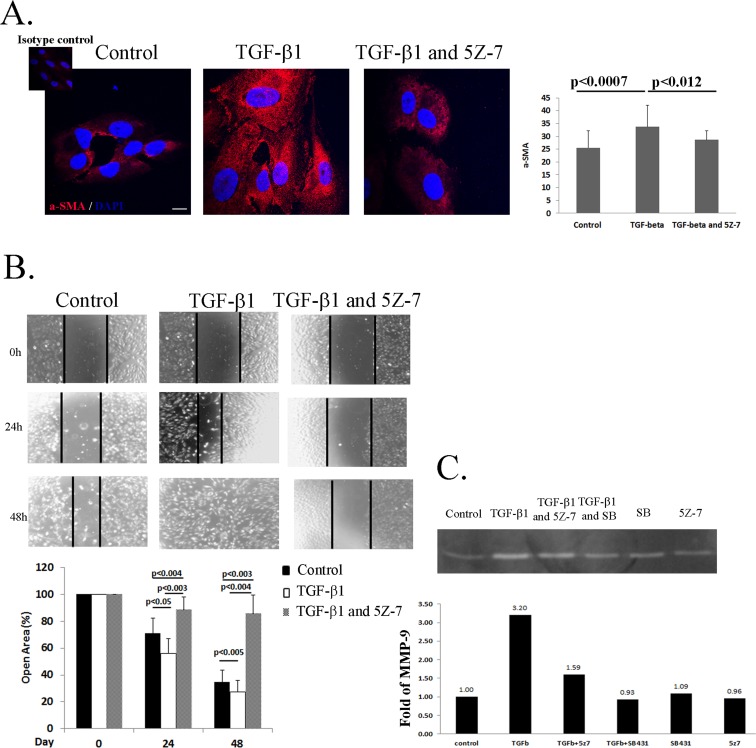
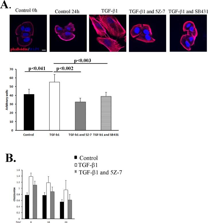
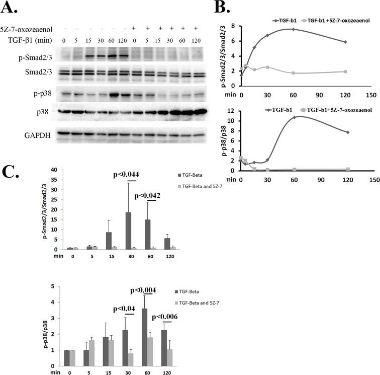
ARPE-19 Cells were treated with 5Z-7 oxozeaenol (TAK1 inhibitor) or SB431542 (TGF-β1 receptor kinase inhibitor) followed by TGF-β1 stimulation. Immunofluorescence, scratch assay Real time PCR and collagen contraction assay assessed the EMT features. The phosphorylation of Smad2/3 and p38 was examined using western blots analysis.
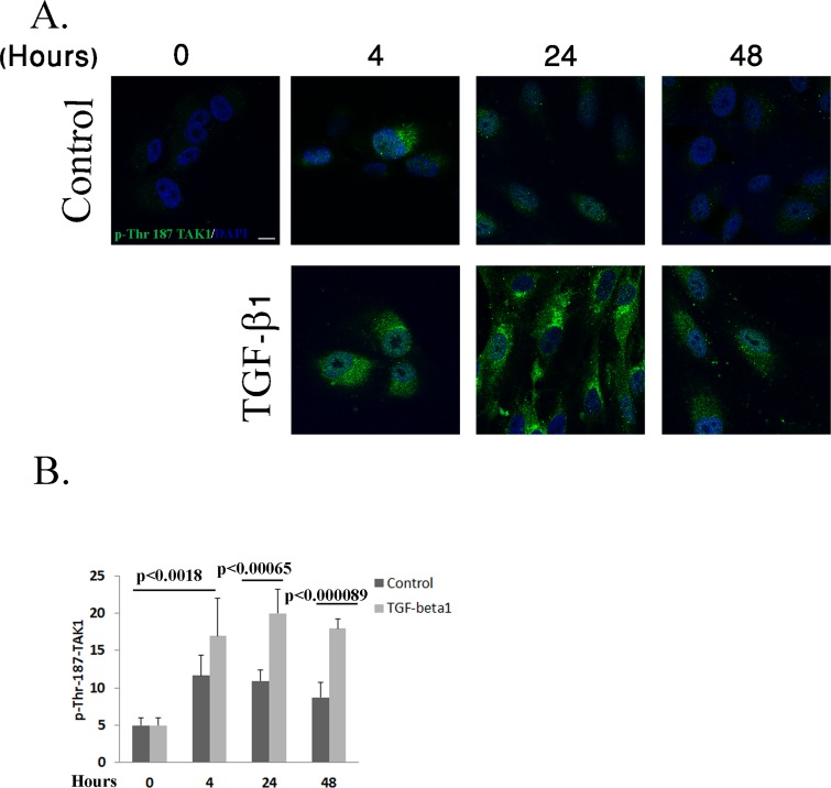
This study demonstrates that stimulation of RPE cells with TGF-β1 increases α-SMA expression, cell migration and cell contractility, all of which are EMT features. Remarkably, addition of TAK1 inhibitor abolishes all these processes. Furthermore, we show hereby that TAK1 regulates not only the activation of the non-canonical cascade of TGF-β1 (p38), but also the canonical cascade, the Smad2/3 activation. Thus, the outcome of the TGF-β response in RPE cells is TAK1 dependent.
CONCLUSIONS/SIGNIFICANCE: This work demonstrated TAK1, a component of the non-canonical pathway of TGF-β1, is a key player in the EMT process, thus provides deep insight into the pathogenesis of PVR. The ability to halt the process of EMT in RPE cells may reduce the severity of the fibrotic response that occurs upon PVR, leading to a better prognosis and increase the probability of success in RD treatment.
增殖性玻璃体视网膜病变(PVR)是一种在视网膜脱离(RD)后作为并发症出现的活跃过程,伴有纤维化组织的形成。PVR期间参与纤维化组织形成的主要细胞是视网膜色素上皮(RPE)细胞。RPE细胞经历上皮-间质转化(EMT),这会导致复杂的视网膜脱离和视力丧失。转化生长因子-β1(TGF-β1)被认为是RPE细胞EMT的主要参与者,尽管其机制尚未完全了解。本研究旨在确定转化生长因子β激活激酶1(TAK1)在RPE细胞EMT过程中可能的作用。
用5Z-7氧代zeaenol(TAK1抑制剂)或SB431542(TGF-β1受体激酶抑制剂)处理ARPE-19细胞,随后进行TGF-β1刺激。通过免疫荧光、划痕试验、实时PCR和胶原收缩试验评估EMT特征。使用蛋白质印迹分析检测Smad2/3和p38的磷酸化。
本研究表明,用TGF-β1刺激RPE细胞会增加α-SMA表达、细胞迁移和细胞收缩性,所有这些都是EMT特征。值得注意的是,添加TAK1抑制剂可消除所有这些过程。此外,我们在此表明TAK1不仅调节TGF-β1的非经典级联反应(p38)的激活,还调节经典级联反应,即Smad2/3的激活。因此,RPE细胞中TGF-β反应的结果是TAK1依赖性的。
结论/意义:这项工作表明TAK1是TGF-β1非经典途径的一个组成部分,是EMT过程中的关键参与者,从而为PVR的发病机制提供了深入见解。阻止RPE细胞中EMT过程的能力可能会降低PVR时发生的纤维化反应的严重程度,从而导致更好的预后并增加RD治疗成功的可能性。