• 文献检索
  • 文档翻译
  • 深度研究
  • 学术资讯
  • Suppr Zotero 插件Zotero 插件
  • 邀请有礼
  • 套餐&价格
  • 历史记录
应用&插件
Suppr Zotero 插件Zotero 插件浏览器插件Mac 客户端Windows 客户端微信小程序
定价
高级版会员购买积分包购买API积分包
服务
文献检索文档翻译深度研究API 文档MCP 服务
关于我们
关于 Suppr公司介绍联系我们用户协议隐私条款
关注我们

Suppr 超能文献

核心技术专利:CN118964589B侵权必究
粤ICP备2023148730 号-1Suppr @ 2026

文献检索

告别复杂PubMed语法,用中文像聊天一样搜索,搜遍4000万医学文献。AI智能推荐,让科研检索更轻松。

立即免费搜索

文件翻译

保留排版,准确专业,支持PDF/Word/PPT等文件格式,支持 12+语言互译。

免费翻译文档

深度研究

AI帮你快速写综述,25分钟生成高质量综述,智能提取关键信息,辅助科研写作。

立即免费体验

雷珠单抗治疗的糖尿病性黄斑水肿患者队列1年随访时的多焦视网膜电图变化

Multifocal electroretinography changes at the 1-year follow-up in a cohort of diabetic macular edema patients treated with ranibizumab.

作者信息

Baget-Bernaldiz Marc, Romero-Aroca Pedro, Bautista-Perez Angel, Mercado Joaquin

机构信息

Ophthalmic Service, University Hospital Sant Joan, Reus, Spain.

Institut de Investigacio Sanitaria Pere Virgili [IISPV], University Rovira and Virgili, Tarragona, Spain.

出版信息

Doc Ophthalmol. 2017 Oct;135(2):85-96. doi: 10.1007/s10633-017-9601-2. Epub 2017 Aug 4.

DOI:10.1007/s10633-017-9601-2
PMID:28779336
原文链接:https://pmc.ncbi.nlm.nih.gov/articles/PMC5606940/
Abstract

PURPOSE

To determine the changes in the multifocal electroretinogram (mfERG) at 1 year in a clinical series of diabetic macular edema (DME) patients treated with ranibizumab (RNBZ) using a pro re nata protocol.

METHODS

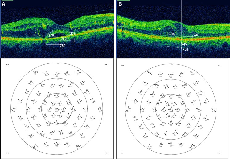
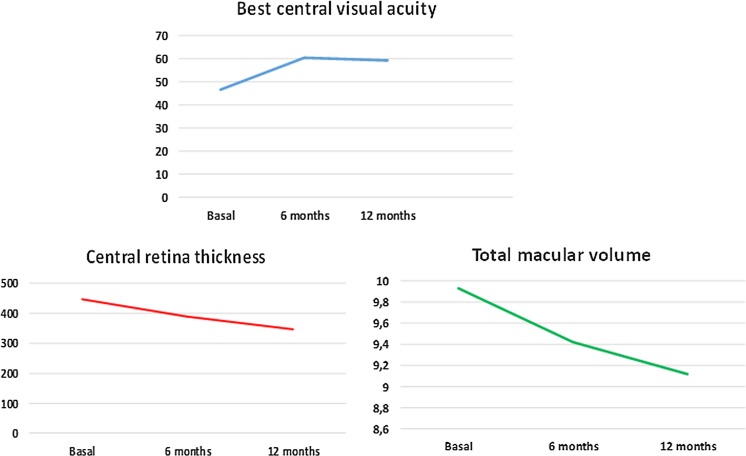
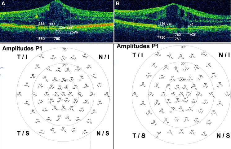
We analyzed a clinical series of 35 eyes of 35 patients with DME at baseline and after treating them with RNBZ over 1 year, in order to determine the change in the macular function, which was assessed by means of the response density and the implicit time of the first-order kernel (FOK) P1 wave of the mfERG at the foveola (R1), fovea (R2) and parafovea (R3). These electrophysiological parameters were studied taking into account different independent variables, such as DME type, degree of diabetic retinopathy (DR), level of preservation of both the ellipsoid zone (IS/OS) and the external limiting membrane (ELM) and changes in central retinal thickness (CRT) and total macular volume (TMV). We also studied the relationship between the response density and the best-corrected visual acuity (BCVA).

RESULTS

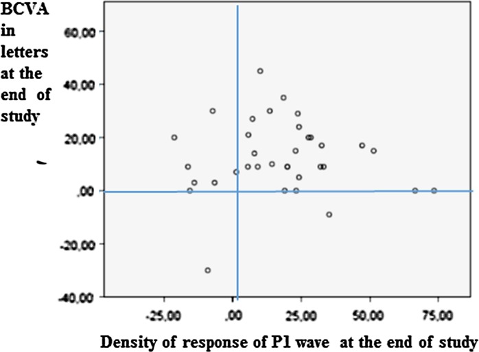
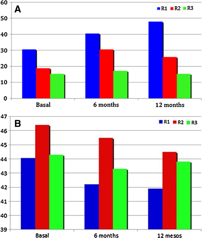
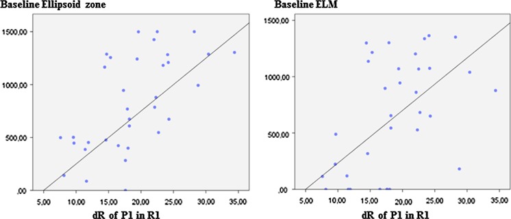
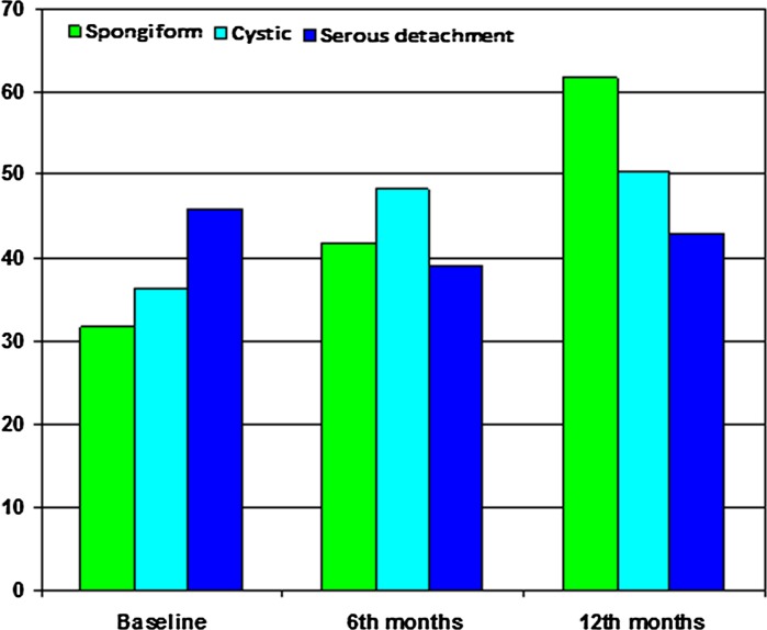
Eyes with cystic and spongiform DME showed better response density with respect to the serous type (p < 0.001) at baseline. Similarly, eyes with high IS/OS and ELM preservation rates showed higher initial response density compared to the others (p < 0.001). Eyes with moderate DR had better response density compared to those with severe and proliferative DR (p = 0.001). At the beginning of the study, those eyes with proliferative and severe DR showed longer implicit times with respect to those with moderate DR (p = 0.04). The response density significantly increased in eyes that anatomically restored the IS/OS and the ELM after being treated with RNBZ (both p < 0.001). Similarly, eyes with spongiform DME further improved the response density with respect to those with cystic and serous DME (p < 0.001). On the contrary, eyes with hard exudates showed less improvement in their response density at the end of the study (p < 0.001). We observed a significant relationship between BCVA and the response density achieved at the end of the study (p = 0.012). Eyes with severe and proliferative DR significantly shortened implicit time compared to those with moderate DR (p = 0.04).

CONCLUSIONS

The multifocal electroretinogram allowed us to differentiate groups of eyes with DME according to their electrophysiological profile, both initially and after being treated with RNBZ. Ranibizumab increased the response density in all DME types included in the study, with a maximum response in those eyes with spongiform type. Once treated with RNBZ, the macular electrophysiological activity improved in eyes that had a well-preserved ellipsoid zone and ELM. The presence of hard exudates was a limitation to the response density achieved at the foveola.

https://cdn.ncbi.nlm.nih.gov/pmc/blobs/6615/5606940/802d8763d9ab/10633_2017_9601_Fig7_HTML.jpg
https://cdn.ncbi.nlm.nih.gov/pmc/blobs/6615/5606940/72a9dace3512/10633_2017_9601_Fig1_HTML.jpg
https://cdn.ncbi.nlm.nih.gov/pmc/blobs/6615/5606940/5634a6d37ccc/10633_2017_9601_Fig2_HTML.jpg
https://cdn.ncbi.nlm.nih.gov/pmc/blobs/6615/5606940/89623155a702/10633_2017_9601_Fig3_HTML.jpg
https://cdn.ncbi.nlm.nih.gov/pmc/blobs/6615/5606940/203b3f01c3ae/10633_2017_9601_Fig4_HTML.jpg
https://cdn.ncbi.nlm.nih.gov/pmc/blobs/6615/5606940/d680bd395ac1/10633_2017_9601_Fig5_HTML.jpg
https://cdn.ncbi.nlm.nih.gov/pmc/blobs/6615/5606940/8131e2abfc33/10633_2017_9601_Fig6_HTML.jpg
https://cdn.ncbi.nlm.nih.gov/pmc/blobs/6615/5606940/802d8763d9ab/10633_2017_9601_Fig7_HTML.jpg
摘要

目的

采用按需给药方案,确定雷珠单抗(RNBZ)治疗的糖尿病性黄斑水肿(DME)患者临床系列中1年时多焦视网膜电图(mfERG)的变化。

方法

我们分析了35例DME患者的35只眼在基线时以及用RNBZ治疗1年后的情况,以确定黄斑功能的变化,通过mfERG在中心凹(R1)、黄斑中心凹(R2)和黄斑旁中心凹(R3)处的一阶核(FOK)P1波的反应密度和隐含时间进行评估。研究这些电生理参数时考虑了不同的独立变量,如DME类型、糖尿病视网膜病变(DR)程度、椭圆体带(IS/OS)和外界膜(ELM)的保存水平以及中心视网膜厚度(CRT)和黄斑总体积(TMV)的变化。我们还研究了反应密度与最佳矫正视力(BCVA)之间的关系。

结果

在基线时,囊样和海绵状DME的眼相对于浆液性类型显示出更好的反应密度(p < 0.001)。同样,IS/OS和ELM保存率高的眼与其他眼相比,初始反应密度更高(p < 0.001)。中度DR的眼与重度和增殖性DR的眼相比,反应密度更好(p = 0.001)。在研究开始时,增殖性和重度DR的眼相对于中度DR的眼显示出更长的隐含时间(p = 0.04)。用RNBZ治疗后解剖学上恢复了IS/OS和ELM的眼中,反应密度显著增加(两者p < 0.001)。同样地,海绵状DME的眼相对于囊样和浆液性DME的眼,反应密度进一步改善(p < 0.001)。相反,有硬性渗出的眼在研究结束时反应密度改善较少(p < 0.001)。我们观察到BCVA与研究结束时达到的反应密度之间存在显著关系(p = 0.012)。重度和增殖性DR的眼与中度DR的眼相比,隐含时间显著缩短(p = 0.04)。

结论

多焦视网膜电图使我们能够根据电生理特征在初始时以及用RNBZ治疗后区分DME的眼组。雷珠单抗增加了研究中包括的所有DME类型的反应密度,在海绵状类型的眼中反应最大。一旦用RNBZ治疗,椭圆体带和ELM保存良好的眼中黄斑电生理活动得到改善。硬性渗出的存在是中心凹处反应密度达到的一个限制因素。

相似文献

1
Multifocal electroretinography changes at the 1-year follow-up in a cohort of diabetic macular edema patients treated with ranibizumab.雷珠单抗治疗的糖尿病性黄斑水肿患者队列1年随访时的多焦视网膜电图变化
Doc Ophthalmol. 2017 Oct;135(2):85-96. doi: 10.1007/s10633-017-9601-2. Epub 2017 Aug 4.
2
Effectiveness of Intravitreal Ranibizumab for Diabetic Macular Edema with Serous Retinal Detachment.玻璃体内注射雷珠单抗治疗伴有浆液性视网膜脱离的糖尿病性黄斑水肿的疗效
Korean J Ophthalmol. 2018 Aug;32(4):296-302. doi: 10.3341/kjo.2017.0117.
3
The macular function and structure in patients with diabetic macular edema before and after ranibizumab treatment.雷珠单抗治疗前后糖尿病性黄斑水肿患者的黄斑功能与结构
Doc Ophthalmol. 2016 Apr;132(2):111-22. doi: 10.1007/s10633-016-9531-4. Epub 2016 Mar 21.
4
Optical Coherence Tomography and Multifocal Electroretinography in Diabetic Macular Edema: A Neurovascular Relation With Vision.光学相干断层扫描与多焦视网膜电图在糖尿病性黄斑水肿中的应用:与视力的神经血管关系
Ophthalmic Surg Lasers Imaging Retina. 2016 Jul 1;47(7):626-31. doi: 10.3928/23258160-20160707-03.
5
Visual and Anatomic Outcomes in Patients with Diabetic Macular Edema with Limited Initial Anatomic Response to Ranibizumab in RIDE and RISE.抗 VEGF 药物治疗糖尿病黄斑水肿初始疗效有限患者的视力和解剖学结局: RIDE 和 RISE 研究
Ophthalmology. 2016 Jun;123(6):1345-50. doi: 10.1016/j.ophtha.2016.02.007. Epub 2016 Mar 15.
6
Structural and functional assessment after intravitreal injection of ranibizumab in diabetic macular edema.玻璃体内注射雷珠单抗治疗糖尿病性黄斑水肿后的结构和功能评估
Doc Ophthalmol. 2017 Dec;135(3):165-173. doi: 10.1007/s10633-017-9604-z. Epub 2017 Jul 29.
7
Effects of intravitreal injection of ranibizumab on choroidal structure and blood flow in eyes with diabetic macular edema.玻璃体内注射雷珠单抗对糖尿病性黄斑水肿患者脉络膜结构和血流的影响。
Graefes Arch Clin Exp Ophthalmol. 2018 May;256(5):885-892. doi: 10.1007/s00417-018-3939-3. Epub 2018 Mar 1.
8
Functional and morphological changes in diabetic macular edema over the course of anti-vascular endothelial growth factor treatment.糖尿病性黄斑水肿在抗血管内皮生长因子治疗过程中的功能和形态变化。
Acta Ophthalmol. 2013 Nov;91(7):e529-36. doi: 10.1111/aos.12153. Epub 2013 May 7.
9
Multifocal electroretinogram in diabetic macular edema and its correlation with different optical coherence tomography features.多焦视网膜电图在糖尿病性黄斑水肿及其与不同光学相干断层扫描特征的相关性。
Int Ophthalmol. 2020 Mar;40(3):571-581. doi: 10.1007/s10792-019-01215-4. Epub 2019 Nov 11.
10
Relationship Between Visual Acuity and Retinal Thickness During Anti-Vascular Endothelial Growth Factor Therapy for Retinal Diseases.视网膜疾病抗血管内皮生长因子治疗期间视力与视网膜厚度的关系
Am J Ophthalmol. 2017 Aug;180:8-17. doi: 10.1016/j.ajo.2017.05.014. Epub 2017 May 24.

引用本文的文献

1
Effects of Dexamethasone Intravitreal Implant on Multifocal Electroretinography in Diabetic Macular Oedema.地塞米松玻璃体内植入物对糖尿病性黄斑水肿多焦视网膜电图的影响
Drug Des Devel Ther. 2024 Nov 25;18:5367-5375. doi: 10.2147/DDDT.S477677. eCollection 2024.
2
Differences in Multifocal Electroretinogram Study in Two Populations of Type 1 and Type 2 Diabetes Mellitus Patients without Diabetic Retinopathy.1型和2型糖尿病无糖尿病视网膜病变患者两组人群的多焦视网膜电图研究差异
J Clin Med. 2022 Sep 30;11(19):5824. doi: 10.3390/jcm11195824.
3
Multifocal Electroretinogram Alterations after Intravitreal Ranibizumab Treatment in Diabetic Macular Edema.

本文引用的文献

1
Global Estimates on the Number of People Blind or Visually Impaired by Diabetic Retinopathy: A Meta-analysis From 1990 to 2010.全球因糖尿病视网膜病变致盲或视力受损人数的估计:1990 年至 2010 年的荟萃分析。
Diabetes Care. 2016 Sep;39(9):1643-9. doi: 10.2337/dc15-2171.
2
The Impact of Systemic Factors on Clinical Response to Ranibizumab for Diabetic Macular Edema.系统性因素对雷珠单抗治疗糖尿病性黄斑水肿临床疗效的影响。
Ophthalmology. 2016 Jul;123(7):1581-7. doi: 10.1016/j.ophtha.2016.03.038. Epub 2016 May 24.
3
Aflibercept, Bevacizumab, or Ranibizumab for Diabetic Macular Edema: Two-Year Results from a Comparative Effectiveness Randomized Clinical Trial.
玻璃体内注射雷珠单抗治疗糖尿病性黄斑水肿后的多焦视网膜电图改变
Beyoglu Eye J. 2022 Feb 18;7(1):39-46. doi: 10.14744/bej.2021.60590. eCollection 2022.
4
Diabetic Retinopathy: From Animal Models to Cellular Signaling.糖尿病视网膜病变:从动物模型到细胞信号转导。
Int J Mol Sci. 2022 Jan 27;23(3):1487. doi: 10.3390/ijms23031487.
5
Assessment of optical coherence tomography angiography and multifocal electroretinography in eyes with and without nonproliferative diabetic retinopathy.评估伴有和不伴有非增生性糖尿病性视网膜病变的眼的光相干断层扫描血管造影和多焦视网膜电图。
Indian J Ophthalmol. 2021 Nov;69(11):3235-3240. doi: 10.4103/ijo.IJO_869_21.
6
Managing Diabetic Macular Edema in Clinical Practice: Systematic Review and Meta-Analysis of Current Strategies and Treatment Options.临床实践中糖尿病性黄斑水肿的管理:当前策略与治疗选择的系统评价和荟萃分析
Clin Ophthalmol. 2021 Jan 29;15:375-385. doi: 10.2147/OPTH.S236423. eCollection 2021.
7
Long-term full-field and multifocal electroretinographic changes after treatment with ranibizumab in patients with diabetic macular edema.长期全视野和多焦视网膜电图在糖尿病黄斑水肿患者接受雷珠单抗治疗后的变化。
Int Ophthalmol. 2021 Apr;41(4):1487-1501. doi: 10.1007/s10792-021-01712-5. Epub 2021 Jan 23.
8
Anatomical and functional outcomes of subthreshold micropulse laser versus intravitreal ranibizumab injection in treatment of diabetic macular edema.阈下微脉冲激光与玻璃体内注射雷珠单抗治疗糖尿病性黄斑水肿的解剖学和功能结局
Int J Retina Vitreous. 2020 Dec 3;6(1):63. doi: 10.1186/s40942-020-00265-6.
9
Efficiency analysis by mfERG and OCT of intravitreal injection with ranibizumab on diabetic macular edema.雷珠单抗玻璃体腔内注射治疗糖尿病性黄斑水肿的多焦视网膜电图和光学相干断层扫描效率分析
Int J Ophthalmol. 2020 Jul 18;13(7):1092-1096. doi: 10.18240/ijo.2020.07.12. eCollection 2020.
10
Microperimetry and mfERG as functional measurements in diabetic macular oedema undergoing intravitreal ranibizumab treatment.微视野计和 mfERG 在接受玻璃体内雷珠单抗治疗的糖尿病性黄斑水肿中的功能测量。
Eye (Lond). 2021 May;35(5):1384-1392. doi: 10.1038/s41433-020-1054-2. Epub 2020 Jul 2.
阿柏西普、贝伐单抗或雷珠单抗治疗糖尿病性黄斑水肿:一项比较疗效随机临床试验的两年结果。
Ophthalmology. 2016 Jun;123(6):1351-9. doi: 10.1016/j.ophtha.2016.02.022. Epub 2016 Feb 27.
4
Morphology and Function over a One-Year Follow Up Period after Intravitreal Dexamethasone Implant (Ozurdex) in Patients with Diabetic Macular Edema.糖尿病性黄斑水肿患者玻璃体内植入地塞米松(Ozurdex)后一年随访期的形态学与功能
PLoS One. 2015 Dec 31;10(12):e0145663. doi: 10.1371/journal.pone.0145663. eCollection 2015.
5
A randomized trial to assess functional and structural effects of ranibizumab versus laser in diabetic macular edema (the LUCIDATE study).一项评估雷珠单抗与激光治疗糖尿病性黄斑水肿(LUCIDATE 研究)的功能和结构效果的随机试验。
Am J Ophthalmol. 2014 May;157(5):960-70. doi: 10.1016/j.ajo.2014.02.019. Epub 2014 Feb 14.
6
Association between perifoveal hyperfluorescence and serous retinal detachment in diabetic macular edema.中心凹旁高荧光与糖尿病性黄斑水肿中浆液性视网膜脱离的相关性。
Ophthalmology. 2013 Dec;120(12):2596-2603. doi: 10.1016/j.ophtha.2013.06.014. Epub 2013 Aug 12.
7
Association of retinal sensitivity to integrity of photoreceptor inner/outer segment junction in patients with diabetic macular edema.糖尿病性黄斑水肿患者视网膜感光性与光感受器内外节连接完整性的相关性。
Ophthalmology. 2013 Jun;120(6):1254-61. doi: 10.1016/j.ophtha.2012.12.003. Epub 2013 Mar 15.
8
RPE barrier breakdown in diabetic retinopathy: seeing is believing.糖尿病性视网膜病变中的视网膜色素上皮屏障破坏:眼见为实。
J Ocul Biol Dis Infor. 2011 Jun;4(1-2):83-92. doi: 10.1007/s12177-011-9068-4. Epub 2011 Dec 31.
9
Association between external limiting membrane status and visual acuity in diabetic macular oedema.糖尿病性黄斑水肿中外膜限制膜状态与视力的关系。
Br J Ophthalmol. 2013 Feb;97(2):228-32. doi: 10.1136/bjophthalmol-2011-301418. Epub 2012 Nov 21.
10
Optical coherence tomographic reflectivity of photoreceptors beneath cystoid spaces in diabetic macular edema.糖尿病性黄斑水肿中囊样间隙下感光细胞的光相干断层扫描反射率。
Invest Ophthalmol Vis Sci. 2012 Mar 21;53(3):1506-11. doi: 10.1167/iovs.11-9231.