Histology and Embryology Research Unit, Department of Experimental and Clinical Medicine, University of Florence, Florence, Italy.
Department of Neuro-Urology, Careggi University Hospital, Florence, Italy.
J Cell Mol Med. 2018 Jan;22(1):195-206. doi: 10.1111/jcmm.13308. Epub 2017 Aug 7.
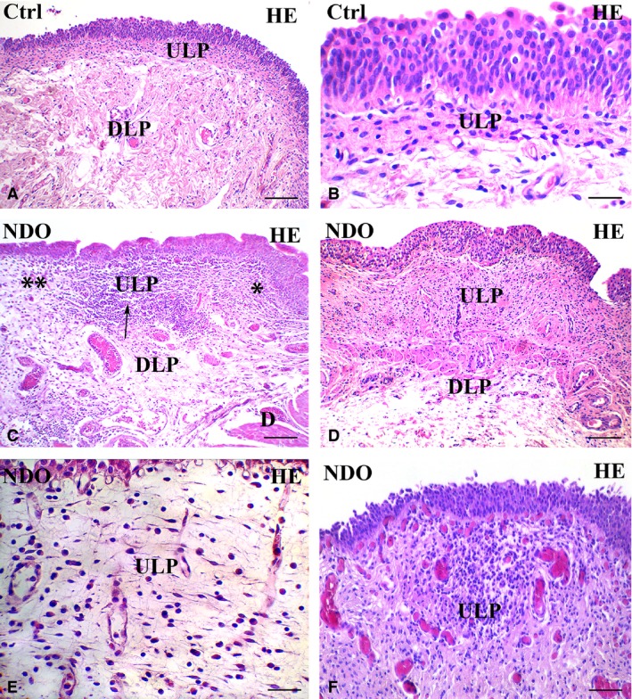
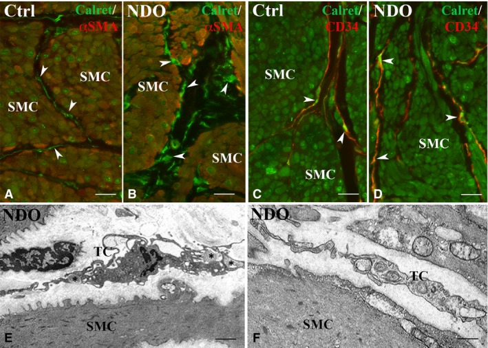
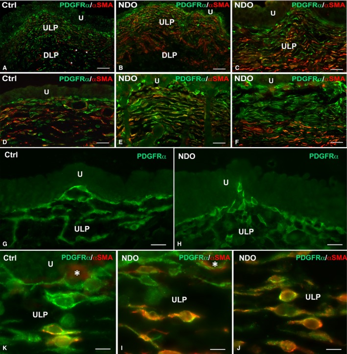
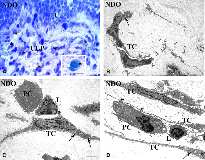
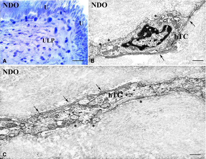
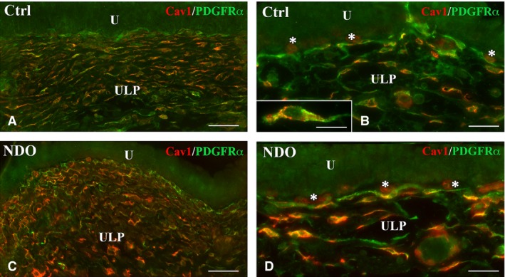
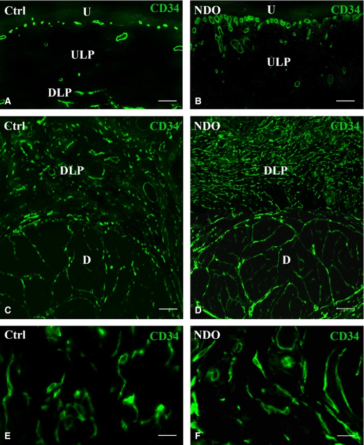
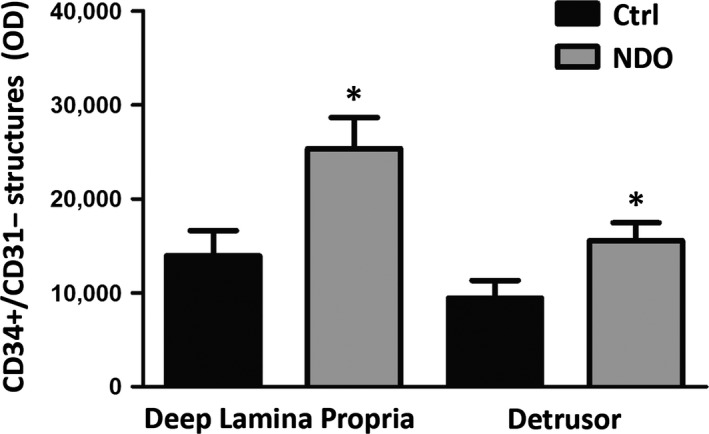
Urinary bladder activity involves central and autonomic nervous systems and bladder wall. Studies on the pathogenesis of voiding disorders such as the neurogenic detrusor overactivity (NDO) due to suprasacral spinal cord lesions have emphasized the importance of an abnormal handling of the afferent signals from urothelium and lamina propria (LP). In the LP (and detrusor), three types of telocytes (TC) are present and form a 3D-network. TC are stromal cells able to form the scaffold that contains and organizes the connective components, to serve as guide for tissue (re)-modelling, to produce trophic and/or regulatory molecules, to share privileged contacts with the immune cells. Specimens of full thickness bladder wall from NDO patients were collected with the aim to investigate possible changes of the three TC types using histology, immunohistochemistry and transmission electron microscopy. The results show that NDO causes several morphological TC changes without cell loss or network interruption. With the exception of those underlying the urothelium, all the TC display signs of activation (increase in Caveolin1 and caveolae, αSMA and thin filaments, Calreticulin and amount of cisternae of the rough endoplasmic reticulum, CD34, euchromatic nuclei and large nucleoli). In all the specimens, a cell infiltrate, mainly consisting in plasma cells located in the vicinity or taking contacts with the TC, is present. In conclusion, our findings show that NDO causes significant changes of all the TC. Notably, these changes can be interpreted as TC adaptability to the pathological condition likely preserving each of their peculiar functions.
膀胱活动涉及中枢和自主神经系统以及膀胱壁。对诸如由于脊髓上损伤引起的逼尿肌过度活动(NDO)等排尿障碍的发病机制的研究强调了异常处理来自尿路上皮和固有层(LP)的传入信号的重要性。在 LP(和逼尿肌)中,存在三种类型的间质细胞(TC)并形成三维网络。TC 是间质细胞,能够形成包含和组织结缔组织成分的支架,作为组织(再)重塑的指导,产生营养和/或调节分子,与免疫细胞共享特权接触。从 NDO 患者的全层膀胱壁采集标本,目的是使用组织学、免疫组织化学和透射电子显微镜研究三种 TC 类型可能发生的变化。结果表明,NDO 导致几种形态 TC 变化,而不会导致细胞丢失或网络中断。除了位于尿路上皮下方的 TC 外,所有 TC 都显示出激活的迹象(Caveolin1 和小窝增加,αSMA 和细纤维,钙网蛋白和粗面内质网的内质网池数量增加,CD34,常染色质核和大核仁)。在所有标本中,存在主要由浆细胞组成的细胞浸润,这些浆细胞位于 TC 附近或与 TC 接触。总之,我们的研究结果表明,NDO 导致所有 TC 发生显著变化。值得注意的是,这些变化可以被解释为 TC 对病理条件的适应性,可能保留了它们各自的特殊功能。