Department of Orthopedics, Guangdong Key Lab of Orthopedic Technology and Implant, Guangzhou General Hospital of Guangzhou Military Command, 111 Liuhua Road, Guangzhou, 510010, China.
Department of Chemistry and Biochemistry, Stephenson Life Sciences Research Center, University of Oklahoma, Norman, OK, 73019, USA.
Sci Rep. 2017 Aug 8;7(1):7626. doi: 10.1038/s41598-017-07243-3.
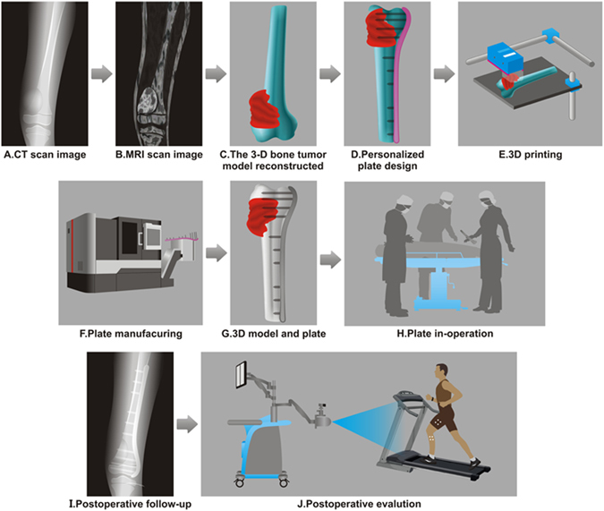
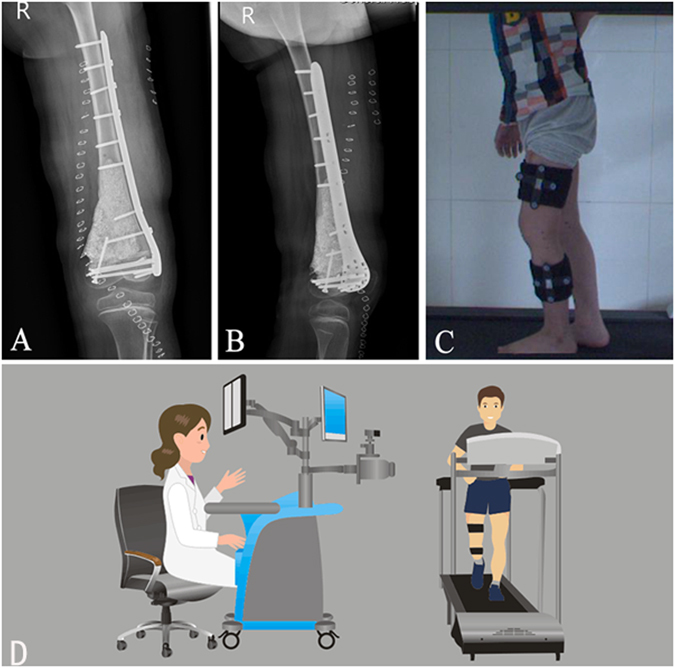
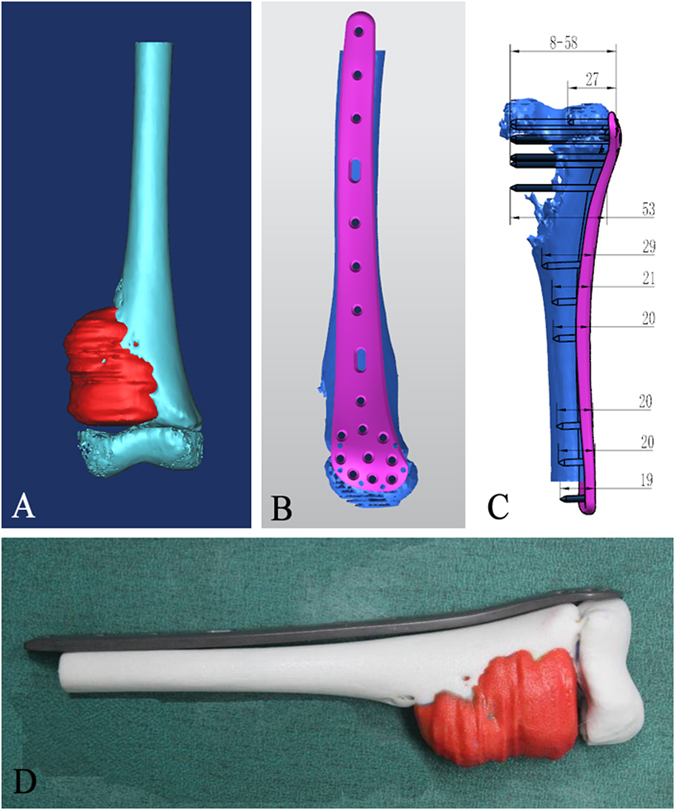
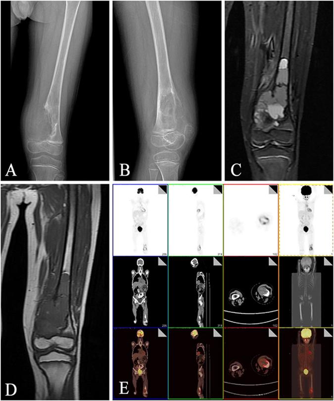
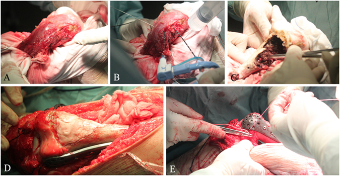
Microwave ablation has been widely accepted in treating bone tumor. However, its procedure is time-consuming and usually results in postoperative fractures. To solve this problem, we designed and fabricated titanium plates customized to the patients' bone structures. The personalized titanium plates were then used for fixation after the removal of tumorous tissue. Specifically, 3D models of tumor-bearing bone segments were constructed by using computed tomography (CT) and magnetic resonance imaging (MRI). The 3D models were used to design the personalized titanium plates. The plate model was transferred into a numerical control machine for manufacturing the personalized titanium plates by 3D printing. The plates were then surgically implanted for reconstruction assistance following microwave-induced hyperthermia to remove the bone tumor. Implementation parameters and knee functions were then evaluated. No postoperative fractures, implant failures or loosening problems occurred; mean Musculoskeletal Tumor Society score was 27.17 from the latest follow-up. Mean maximum flexion of affected knees was 114.08°. The results of knee gait analysis were comparable with normal population data. Our work suggests that personalized titanium plates can significantly improve the clinical outcomes in the surgical removal of bone tumor. This study represents the first-time effort in using personalized titanium plates for such surgery.
微波消融术已被广泛应用于治疗骨肿瘤。然而,其过程耗时且常导致术后骨折。为了解决这个问题,我们设计并制造了定制化的钛板,以适应患者的骨骼结构。在切除肿瘤组织后,这些个性化钛板用于固定。具体来说,通过计算机断层扫描(CT)和磁共振成像(MRI)构建载瘤骨段的 3D 模型。使用这些 3D 模型来设计个性化钛板。将板模型传输到数控机床,通过 3D 打印制造个性化钛板。然后,通过微波诱导高热切除骨肿瘤,将这些钛板进行手术植入以辅助重建。接下来评估实施参数和膝关节功能。没有发生术后骨折、植入物失败或松动的问题;末次随访的肌肉骨骼肿瘤学会评分平均为 27.17 分。受影响膝关节的最大屈曲平均值为 114.08°。膝关节步态分析的结果与正常人群数据相当。我们的工作表明,个性化钛板可显著改善骨肿瘤切除手术的临床效果。本研究代表了首次在该类手术中使用个性化钛板。