• 文献检索
  • 文档翻译
  • 深度研究
  • 学术资讯
  • Suppr Zotero 插件Zotero 插件
  • 邀请有礼
  • 套餐&价格
  • 历史记录
应用&插件
Suppr Zotero 插件Zotero 插件浏览器插件Mac 客户端Windows 客户端微信小程序
定价
高级版会员购买积分包购买API积分包
服务
文献检索文档翻译深度研究API 文档MCP 服务
关于我们
关于 Suppr公司介绍联系我们用户协议隐私条款
关注我们

Suppr 超能文献

核心技术专利:CN118964589B侵权必究
粤ICP备2023148730 号-1Suppr @ 2026

文献检索

告别复杂PubMed语法,用中文像聊天一样搜索,搜遍4000万医学文献。AI智能推荐,让科研检索更轻松。

立即免费搜索

文件翻译

保留排版,准确专业,支持PDF/Word/PPT等文件格式,支持 12+语言互译。

免费翻译文档

深度研究

AI帮你快速写综述,25分钟生成高质量综述,智能提取关键信息,辅助科研写作。

立即免费体验

单向多孔羟基磷灰石骨替代物在皮质骨缺损部位的骨再生与重塑:植入后一年和两年的组织学分析

Bone Regeneration and Remodeling within a Unidirectional Porous Hydroxyapatite Bone Substitute at a Cortical Bone Defect Site: Histological Analysis at One and Two Years after Implantation.

作者信息

Iwasashi Masashi, Funayama Toru, Watanabe Arata, Noguchi Hiroshi, Tsukanishi Toshinori, Suetsugu Yasushi, Makihara Takeshi, Ochiai Naoyuki, Yamazaki Masashi, Sakane Masataka

机构信息

Department of Orthopaedic Surgery, Tsukuba Medical Center Hospital, 1-3-1 Amakubo, Tsukuba, Ibaraki 305-8550, Japan.

Department of Orthopaedic Surgery, Kenpoku Medical Center, Takahagi Kyodo Hospital, 1006-9 Agehocho, Kamitetsuna, Takahagi, Ibaraki 318-0004, Japan.

出版信息

Materials (Basel). 2015 Jul 30;8(8):4884-4894. doi: 10.3390/ma8084884.

DOI:10.3390/ma8084884
PMID:28793479
原文链接:https://pmc.ncbi.nlm.nih.gov/articles/PMC5455501/
Abstract

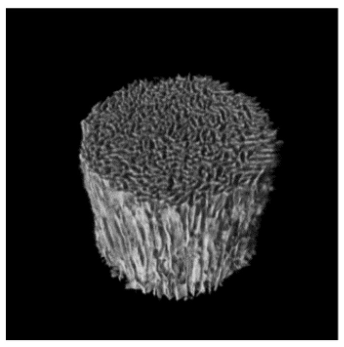
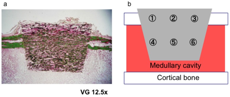
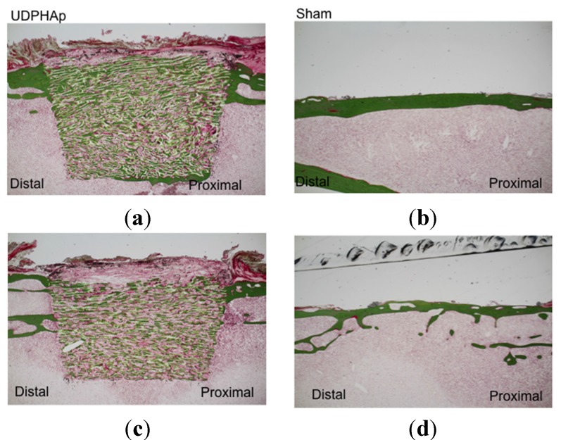
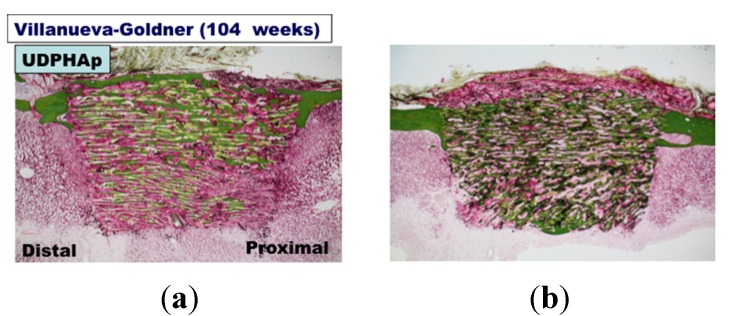
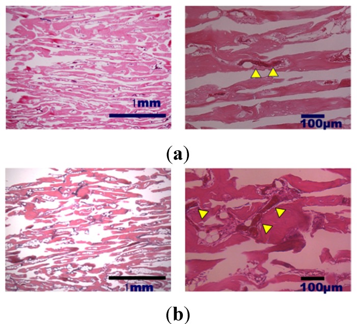
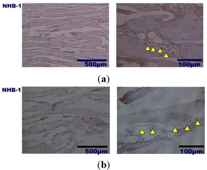
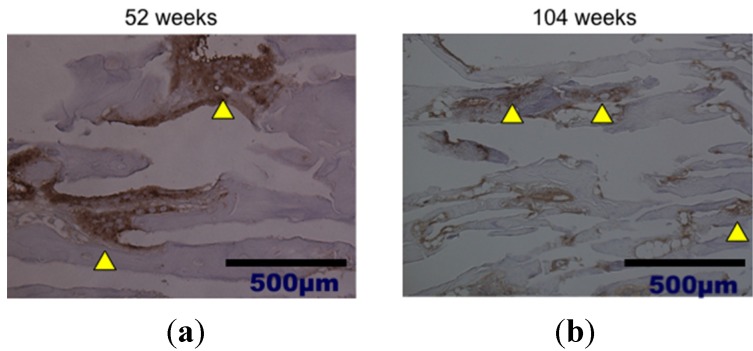
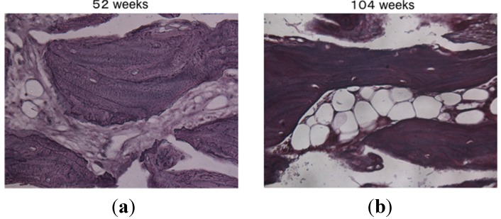
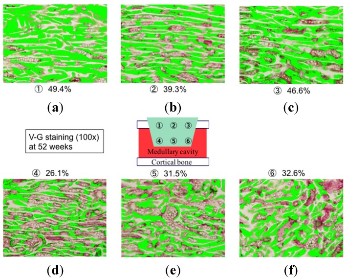
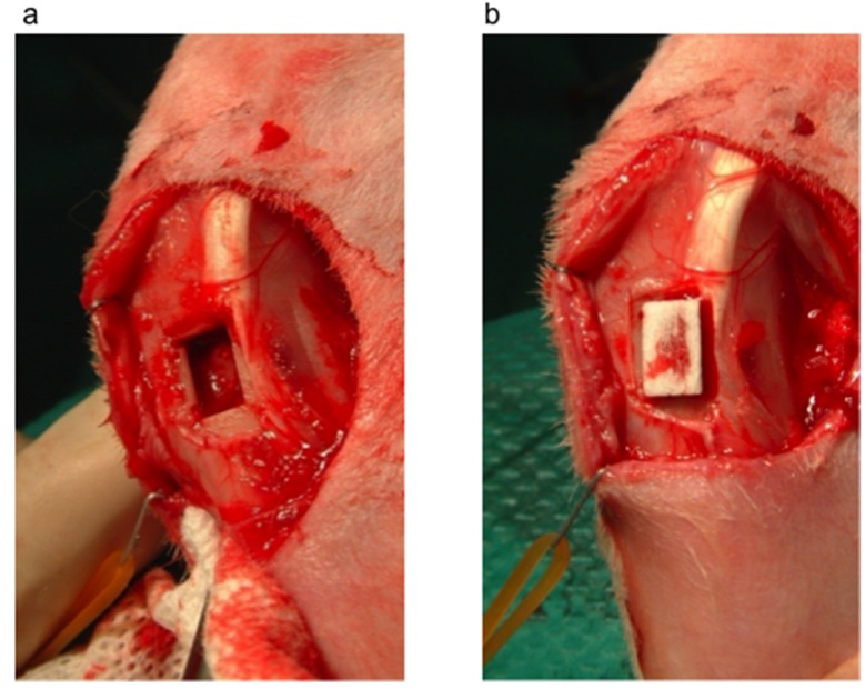
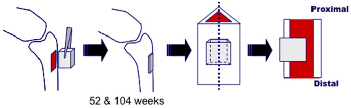
Unidirectional porous hydroxyapatite (UDPHAp) is an artificial bone substitute with a unique microstructure consisting of 100-300-µm oval pores that present the material unidirectionally. UDPHAp has a compression strength of 14 MPa and a porosity of 75%, which promotes cell migration and capillary formation within the material. Despite these advantageous properties, bone remodeling and bone formation with UDPHAp remain unclear. To examine long-term remodeling and differences in bone formation based on the defect site, trapezoidal prism-shaped UDPHAp blocks were implanted into rectangular-shaped cortical bone defects in the proximal tibia of Japanese white rabbits. Histological analysis performed at 52 and 104 weeks after implantation revealed that bone and capillaries had formed within the implanted UDPHAp material. Bone formed within the UDPHAp implanted in the cortical defect of rabbit tibia and remodel up to two years. The percentage of new bone area within UDPHAp was larger in cortical lesions than that in medullary lesions. These findings suggest that UDPHAp is a promising material for the repair of non-critical-sized cortical bone defects.

摘要

单向多孔羟基磷灰石(UDPHAp)是一种人工骨替代物,具有独特的微观结构,由100 - 300微米的椭圆形孔隙组成,这些孔隙使材料呈现单向性。UDPHAp的抗压强度为14兆帕,孔隙率为75%,这促进了材料内细胞迁移和毛细血管形成。尽管具有这些有利特性,但UDPHAp的骨重塑和骨形成情况仍不清楚。为了研究基于缺损部位的长期重塑和骨形成差异,将梯形棱柱形UDPHAp块植入日本白兔胫骨近端的矩形皮质骨缺损处。植入后52周和104周进行的组织学分析显示,植入的UDPHAp材料内已形成骨和毛细血管。植入兔胫骨皮质缺损处的UDPHAp内形成的骨可重塑长达两年。UDPHAp内新骨面积的百分比在皮质病变中比在髓质病变中更大。这些发现表明,UDPHAp是修复非临界尺寸皮质骨缺损的一种有前景的材料。

https://cdn.ncbi.nlm.nih.gov/pmc/blobs/19ab/5455501/f4b45850f410/materials-08-04884-g011.jpg
https://cdn.ncbi.nlm.nih.gov/pmc/blobs/19ab/5455501/b941ed7f670c/materials-08-04884-g001.jpg
https://cdn.ncbi.nlm.nih.gov/pmc/blobs/19ab/5455501/65167be666f8/materials-08-04884-g002.jpg
https://cdn.ncbi.nlm.nih.gov/pmc/blobs/19ab/5455501/129e064256bd/materials-08-04884-g003a.jpg
https://cdn.ncbi.nlm.nih.gov/pmc/blobs/19ab/5455501/6c38efc3c0f0/materials-08-04884-g004.jpg
https://cdn.ncbi.nlm.nih.gov/pmc/blobs/19ab/5455501/968de163aba7/materials-08-04884-g005.jpg
https://cdn.ncbi.nlm.nih.gov/pmc/blobs/19ab/5455501/9aef4fdade89/materials-08-04884-g006.jpg
https://cdn.ncbi.nlm.nih.gov/pmc/blobs/19ab/5455501/a09119c4d62e/materials-08-04884-g007.jpg
https://cdn.ncbi.nlm.nih.gov/pmc/blobs/19ab/5455501/21490362f919/materials-08-04884-g008.jpg
https://cdn.ncbi.nlm.nih.gov/pmc/blobs/19ab/5455501/4575e1557a42/materials-08-04884-g009.jpg
https://cdn.ncbi.nlm.nih.gov/pmc/blobs/19ab/5455501/2a1f58ad0bfe/materials-08-04884-g010.jpg
https://cdn.ncbi.nlm.nih.gov/pmc/blobs/19ab/5455501/f4b45850f410/materials-08-04884-g011.jpg

相似文献

1
Bone Regeneration and Remodeling within a Unidirectional Porous Hydroxyapatite Bone Substitute at a Cortical Bone Defect Site: Histological Analysis at One and Two Years after Implantation.单向多孔羟基磷灰石骨替代物在皮质骨缺损部位的骨再生与重塑:植入后一年和两年的组织学分析
Materials (Basel). 2015 Jul 30;8(8):4884-4894. doi: 10.3390/ma8084884.
2
Formation of osteon-like structures in unidirectional porous hydroxyapatite substitute.在各向异性多孔羟基磷灰石替代物中形成骨单位样结构。
J Biomed Mater Res B Appl Biomater. 2018 Oct;106(7):2665-2672. doi: 10.1002/jbm.b.34083. Epub 2018 Feb 13.
3
Clinical Application of Unidirectional Porous Hydroxyapatite to Bone Tumor Surgery and Other Orthopedic Surgery.单向多孔羟基磷灰石在骨肿瘤手术及其他骨科手术中的临床应用
Biomimetics (Basel). 2024 May 15;9(5):294. doi: 10.3390/biomimetics9050294.
4
Physico-Chemical, In Vitro, and In Vivo Evaluation of a 3D Unidirectional Porous Hydroxyapatite Scaffold for Bone Regeneration.用于骨再生的3D单向多孔羟基磷灰石支架的物理化学、体外和体内评价
Materials (Basel). 2017 Jan 3;10(1):33. doi: 10.3390/ma10010033.
5
Radiographic and clinical assessment of unidirectional porous hydroxyapatite to treat benign bone tumors.单向多孔羟基磷灰石治疗良性骨肿瘤的影像学与临床评估
Sci Rep. 2020 Dec 9;10(1):21578. doi: 10.1038/s41598-020-78409-9.
6
Regeneration of the Fibula with Unidirectional Porous Hydroxyapatite.单向多孔羟基磷灰石促进腓骨再生
Case Rep Orthop. 2019 Oct 13;2019:9024643. doi: 10.1155/2019/9024643. eCollection 2019.
7
Novel unidirectional porous hydroxyapatite used as a bone substitute for open wedge high tibial osteotomy.新型单向多孔羟基磷灰石用作开放性楔形高位胫骨截骨术的骨替代物。
J Mater Sci Mater Med. 2014 Nov;25(11):2541-7. doi: 10.1007/s10856-014-5266-5. Epub 2014 Jul 5.
8
Angiogenesis and new bone formation in novel unidirectional porous beta-tricalcium phosphate: a histological study.新型单向多孔β-磷酸三钙中的血管生成与新骨形成:一项组织学研究。
J Artif Organs. 2019 Dec;22(4):294-299. doi: 10.1007/s10047-019-01120-8. Epub 2019 Jul 19.
9
Effects of pore size and implant volume of porous hydroxyapatite/collagen (HAp/Col) on bone formation in a rabbit bone defect model.多孔羟基磷灰石/胶原蛋白(HAp/Col)的孔径和植入体积对兔骨缺损模型中骨形成的影响。
J Med Dent Sci. 2008 Mar;55(1):91-9.
10
Bone neoformation of a novel porous resorbable Si-Ca-P-based ceramic with osteoconductive properties: physical and mechanical characterization, histological and histomorphometric study.具有骨传导特性的新型多孔可吸收硅钙磷基陶瓷的骨新形成:物理和力学表征、组织学和组织形态计量学研究。
Clin Oral Implants Res. 2016 Nov;27(11):1368-1375. doi: 10.1111/clr.12745. Epub 2016 Jan 18.

引用本文的文献

1
Clinical Application of Unidirectional Porous Hydroxyapatite to Bone Tumor Surgery and Other Orthopedic Surgery.单向多孔羟基磷灰石在骨肿瘤手术及其他骨科手术中的临床应用
Biomimetics (Basel). 2024 May 15;9(5):294. doi: 10.3390/biomimetics9050294.
2
Unidirectional porous beta-tricalcium phosphate and hydroxyapatite artificial bone: a review of experimental evaluations and clinical applications.单向多孔β-磷酸三钙和羟基磷灰石人工骨:实验评价和临床应用的综述。
J Artif Organs. 2021 Jun;24(2):103-110. doi: 10.1007/s10047-021-01270-8. Epub 2021 Apr 23.
3
How much platelet-rich plasma can be soak-loaded onto beta-tricalcium phosphate? A comparison with or without a unidirectional porous structure.

本文引用的文献

1
Monocytes induce STAT3 activation in human mesenchymal stem cells to promote osteoblast formation.单核细胞诱导人骨髓间充质干细胞中 STAT3 的激活,促进成骨细胞的形成。
PLoS One. 2012;7(7):e39871. doi: 10.1371/journal.pone.0039871. Epub 2012 Jul 3.
2
Additive manufacturing of wet-spun polymeric scaffolds for bone tissue engineering.用于骨组织工程的湿法纺丝聚合物支架的增材制造。
Biomed Microdevices. 2012 Dec;14(6):1115-27. doi: 10.1007/s10544-012-9677-0.
3
Induction of osteogenesis in mesenchymal stem cells by activated monocytes/macrophages depends on oncostatin M signaling.
β-磷酸三钙能负载多少富血小板血浆?有或无单向多孔结构的比较。
J Rural Med. 2021 Jan;16(1):14-21. doi: 10.2185/jrm.2020-034. Epub 2021 Jan 5.
4
Radiographic and clinical assessment of unidirectional porous hydroxyapatite to treat benign bone tumors.单向多孔羟基磷灰石治疗良性骨肿瘤的影像学与临床评估
Sci Rep. 2020 Dec 9;10(1):21578. doi: 10.1038/s41598-020-78409-9.
5
An overview of de novo bone generation in animal models.动物模型中新骨生成的概述。
J Orthop Res. 2021 Jan;39(1):7-21. doi: 10.1002/jor.24852. Epub 2020 Sep 23.
6
The effect of housing environment on bone healing in a critical radius defect in New Zealand White rabbits.新西兰大白兔临界半径缺损模型中住房环境对骨愈合的影响。
PLoS One. 2020 May 21;15(5):e0233530. doi: 10.1371/journal.pone.0233530. eCollection 2020.
7
Regeneration of the Fibula with Unidirectional Porous Hydroxyapatite.单向多孔羟基磷灰石促进腓骨再生
Case Rep Orthop. 2019 Oct 13;2019:9024643. doi: 10.1155/2019/9024643. eCollection 2019.
8
Angiogenesis and new bone formation in novel unidirectional porous beta-tricalcium phosphate: a histological study.新型单向多孔β-磷酸三钙中的血管生成与新骨形成:一项组织学研究。
J Artif Organs. 2019 Dec;22(4):294-299. doi: 10.1007/s10047-019-01120-8. Epub 2019 Jul 19.
9
Surgical Repair of Acetabular Fracture Using Unidirectional Porous -Tricalcium Phosphate.使用单向多孔磷酸三钙进行髋臼骨折的手术修复
Case Rep Orthop. 2019 Apr 16;2019:6860591. doi: 10.1155/2019/6860591. eCollection 2019.
10
Formation of osteon-like structures in unidirectional porous hydroxyapatite substitute.在各向异性多孔羟基磷灰石替代物中形成骨单位样结构。
J Biomed Mater Res B Appl Biomater. 2018 Oct;106(7):2665-2672. doi: 10.1002/jbm.b.34083. Epub 2018 Feb 13.
激活的单核细胞/巨噬细胞诱导间充质干细胞成骨作用依赖于抑瘤素 M 信号。
Stem Cells. 2012 Apr;30(4):762-72. doi: 10.1002/stem.1040.
4
Coupling of osteogenesis and angiogenesis in bone substitute healing - a brief overview.骨替代物愈合中的成骨与血管生成偶联——简要综述。
Ann Anat. 2012 Mar 20;194(2):171-3. doi: 10.1016/j.aanat.2011.10.002. Epub 2011 Oct 14.
5
Novel fully interconnected porous hydroxyapatite ceramic in surgical treatment of benign bone tumor.新型全互连多孔羟基磷灰石陶瓷在良性骨肿瘤手术治疗中的应用
J Orthop Sci. 2010 Jul;15(4):560-8. doi: 10.1007/s00776-010-1479-8. Epub 2010 Aug 19.
6
A comparative assessment of synthetic ceramic bone substitutes with different composition and microstructure in rabbit femoral condyle model.不同成分和微观结构的合成陶瓷骨替代物在兔股骨髁模型中的比较评估。
J Biomed Mater Res B Appl Biomater. 2009 Nov;91(2):788-798. doi: 10.1002/jbm.b.31457.
7
The biodegradation of hydroxyapatite bone graft substitutes in vivo.羟基磷灰石骨移植替代物在体内的生物降解
Folia Morphol (Warsz). 2006 Feb;65(1):43-8.
8
Porosity of 3D biomaterial scaffolds and osteogenesis.3D生物材料支架的孔隙率与骨生成
Biomaterials. 2005 Sep;26(27):5474-91. doi: 10.1016/j.biomaterials.2005.02.002.
9
Calcium hydroxyapatite ceramic implants in bone tumour surgery. A long-term follow-up study.骨肿瘤手术中羟基磷灰石钙陶瓷植入物。一项长期随访研究。
J Bone Joint Surg Br. 2004 Jul;86(5):719-25. doi: 10.1302/0301-620x.86b5.14242.
10
Osteoconduction at porous hydroxyapatite with various pore configurations.具有不同孔隙结构的多孔羟基磷灰石的骨传导性。
Biomaterials. 2000 Jun;21(12):1291-8. doi: 10.1016/s0142-9612(00)00030-2.