Belotto Renata A, Chavantes Maria Cristina, Tardivo João Paulo, Euzébio Dos Santos Roberto, Fernandes Raquel Civolani Marques, Horliana Anna Carolina Ratto Tempestini, Pavani Christiane, Teixeira da Silva Daniela Fátima
Postgraduate Program in Biophotonics Applied to Health Sciences, Nove de Julho University/UNINOVE, 249 Vergueiro Street, Liberdade, São Paulo, SP, 01504-001, Brazil.
Pérola Byington Hospital, 683 Brig. Luís Antônio Avenue, Bela Vista, São Paulo, SP, 01318-000, Brazil.
BMC Womens Health. 2017 Aug 10;17(1):61. doi: 10.1186/s12905-017-0414-y.
Vulvar lichen sclerosus (VLS) is a lymphocyte-mediated disease of unknown etiology that can cause intense itching as well stenosis, hindering the evacuation and urination. It can also limit the sex life due to severe local pruritus, pain and dyspareunia (pain during sexual intercourse). The standard treatment for this disease is the use of topical corticosteroids to reduce the clinical symptoms and to try to increase disease-free intervals. Photodynamic therapy (PDT), a treatment that associates a light radiation with a photosensitizing agent and photobiomodulation (PBM) are therapies that can promote effective immunomodulatory responses at the application site by means of photophysical and photochemical phenomena from the molecular to the systemic level, which promote their use in chronic dermatoses. The aim is to compare the effects of PDT, PBM, and topical corticosteroid in VLS evaluating clinical, histological, immunohistochemical and spectroscopic responses.
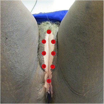
The study is prospective, randomized and controlled, in a population of 60 women with histological diagnoses of VLS. There will be 3 treatments groups: PDT, PBM and topical corticosteroid (control group), where will be allocated by randomization 20 patients in each one. The clinical course will be monitored by measuring local temperature, itching, atrophy, and the area of the lesion. Histologically, the slides will be classified and will have the ordering of collagen fibers quantified. Immunohistochemical analysis will be done using the markers IFN-γ, TGF-β, CD4, CD8, IL-1, p53 and Ki-67. Finally, the spectroscopic evaluation will be done by reflectance. Descriptive and inferential statistical analyses will be conducted to compare the groups and make associations between different responses. The study is an open-label for patients with active symptomatic disease with a period of 1 year follow-up to determine the rate of recurrence in each groups.
The immunological effects of PDT and PBM are described by several authors in inflammatory skin diseases, stimulating the production and organization of the associated collagen. Thus, it is reasonable to determine the efficacy and safety of these new treatments in VLS, in comparison to the control group, analyzing the recurrence time, the impact on the optical properties of the skin, and the benefit to patients.
ClinicalTrials.gov: NCT02416531 .
外阴硬化性苔藓(VLS)是一种病因不明的淋巴细胞介导的疾病,可导致剧烈瘙痒以及狭窄,阻碍排便和排尿。由于严重的局部瘙痒、疼痛和性交困难(性交时疼痛),它还会限制性生活。该病的标准治疗方法是使用外用皮质类固醇来减轻临床症状,并试图延长无病间期。光动力疗法(PDT)是一种将光辐射与光敏剂相结合的治疗方法,光生物调节(PBM)则是通过分子水平到系统水平的光物理和光化学现象,在应用部位促进有效免疫调节反应的疗法,这促使它们被用于慢性皮肤病。目的是比较PDT、PBM和外用皮质类固醇在VLS中的效果,评估临床、组织学、免疫组织化学和光谱学反应。
该研究为前瞻性、随机对照研究,研究对象为60名经组织学诊断为VLS的女性。将有3个治疗组:PDT组、PBM组和外用皮质类固醇组(对照组),每组随机分配20名患者。通过测量局部温度、瘙痒、萎缩和病变面积来监测临床病程。在组织学上,对切片进行分类,并对胶原纤维的排列进行定量。免疫组织化学分析将使用IFN-γ、TGF-β、CD4、CD8、IL-1、p53和Ki-67标记物进行。最后,通过反射率进行光谱评估。将进行描述性和推断性统计分析,以比较各组并建立不同反应之间的关联。该研究对有活动性症状疾病的患者采用开放标签,随访1年以确定各组的复发率。
几位作者在炎症性皮肤病中描述了PDT和PBM的免疫作用,刺激相关胶原的产生和组织。因此,与对照组相比,通过分析复发时间、对皮肤光学性质的影响以及对患者的益处,确定这些新治疗方法在VLS中的疗效和安全性是合理的。
ClinicalTrials.gov:NCT02416531 。