Lu Kun-Han, Cao Jiayue, Oleson Steven Thomas, Powley Terry L, Liu Zhongming
School of Electrical and Computer Engineering and Purdue Institute for Integrative NeurosciencePurdue University.
Weldon School of Biomedical Engineering and Purdue Institute for Integrative NeurosciencePurdue University.
IEEE Trans Biomed Eng. 2017 Nov;64(11):2546-2554. doi: 10.1109/TBME.2017.2737559.
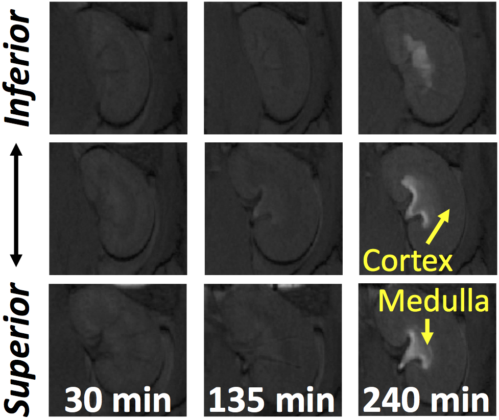
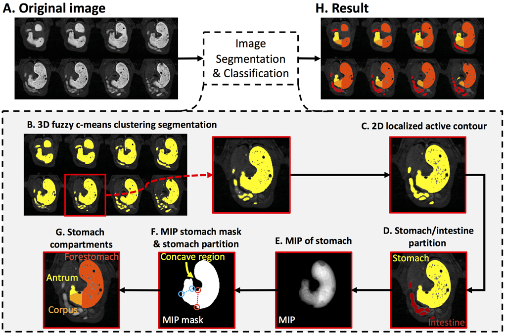
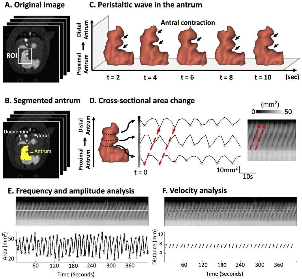
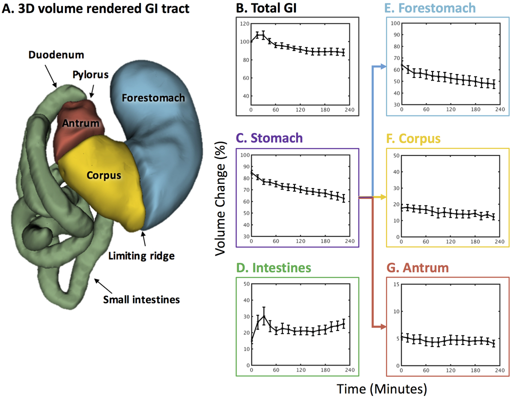
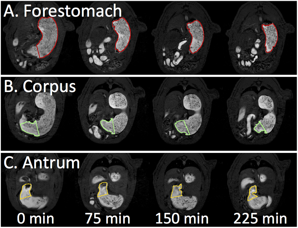
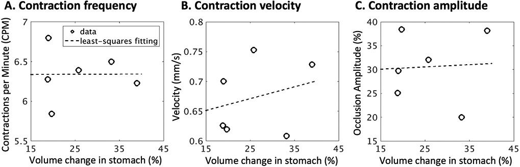
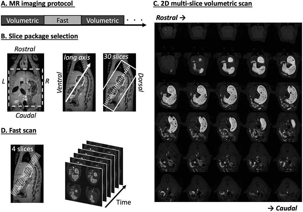
The assessment of gastric emptying and motility in humans and animals typically requires radioactive imaging or invasive measurements. Here, we developed a robust strategy to image and characterize gastric emptying and motility in rats based on contrast-enhanced magnetic resonance imaging (MRI) and computer-assisted image processing. The animals were trained to naturally consume a gadolinium-labeled dietgel while bypassing any need for oral gavage. Following this test meal, the animals were scanned under low-dose anesthesia for high-resolution T1-weighted MRI in 7 Tesla, visualizing the time-varying distribution of the meal with greatly enhanced contrast against non-gastrointestinal (GI) tissues. Such contrast-enhanced images not only depicted the gastric anatomy, but also captured and quantified stomach emptying, intestinal filling, antral contraction, and intestinal absorption with fully automated image processing. Over four postingestion hours, the stomach emptied by 27%, largely attributed to the emptying of the forestomach rather than the corpus and the antrum, and most notable during the first 30 min. Stomach emptying was accompanied by intestinal filling for the first 2 h, whereas afterward intestinal absorption was observable as cumulative contrast enhancement in the renal medulla. The antral contraction was captured as a peristaltic wave propagating from the proximal to distal antrum. The frequency, velocity, and amplitude of the antral contraction were on average 6.34 ± 0.07 contractions per minute, 0.67 ± 0.01 mm/s, and 30.58 ± 1.03%, respectively. These results demonstrate an optimized MRI-based strategy to assess gastric emptying and motility in healthy rats, paving the way for using this technique to understand GI diseases, or test new therapeutics in rat models.The assessment of gastric emptying and motility in humans and animals typically requires radioactive imaging or invasive measurements. Here, we developed a robust strategy to image and characterize gastric emptying and motility in rats based on contrast-enhanced magnetic resonance imaging (MRI) and computer-assisted image processing. The animals were trained to naturally consume a gadolinium-labeled dietgel while bypassing any need for oral gavage. Following this test meal, the animals were scanned under low-dose anesthesia for high-resolution T1-weighted MRI in 7 Tesla, visualizing the time-varying distribution of the meal with greatly enhanced contrast against non-gastrointestinal (GI) tissues. Such contrast-enhanced images not only depicted the gastric anatomy, but also captured and quantified stomach emptying, intestinal filling, antral contraction, and intestinal absorption with fully automated image processing. Over four postingestion hours, the stomach emptied by 27%, largely attributed to the emptying of the forestomach rather than the corpus and the antrum, and most notable during the first 30 min. Stomach emptying was accompanied by intestinal filling for the first 2 h, whereas afterward intestinal absorption was observable as cumulative contrast enhancement in the renal medulla. The antral contraction was captured as a peristaltic wave propagating from the proximal to distal antrum. The frequency, velocity, and amplitude of the antral contraction were on average 6.34 ± 0.07 contractions per minute, 0.67 ± 0.01 mm/s, and 30.58 ± 1.03%, respectively. These results demonstrate an optimized MRI-based strategy to assess gastric emptying and motility in healthy rats, paving the way for using this technique to understand GI diseases, or test new therapeutics in rat models.
对人和动物胃排空及动力的评估通常需要放射性成像或侵入性测量。在此,我们基于对比增强磁共振成像(MRI)和计算机辅助图像处理,开发了一种用于对大鼠胃排空和动力进行成像及特征描述的可靠策略。训练动物自然食用钆标记的饮食凝胶,从而无需进行灌胃。在食用此测试餐之后,在低剂量麻醉下对动物进行扫描,以在7特斯拉磁场中进行高分辨率T1加权MRI,可视化该餐食随时间变化的分布情况,与非胃肠道(GI)组织形成极大增强的对比。这种对比增强图像不仅描绘了胃的解剖结构,还通过全自动图像处理捕获并量化了胃排空、肠道充盈、胃窦收缩和肠道吸收情况。在摄入食物后的四个小时内,胃排空了27%,这主要归因于前胃的排空,而非胃体和胃窦,且在最初30分钟最为明显。在最初2小时内,胃排空伴随着肠道充盈,而此后可观察到肠道吸收表现为肾髓质中对比剂的累积增强。胃窦收缩表现为从胃窦近端向远端传播的蠕动波。胃窦收缩的频率、速度和幅度平均分别为每分钟6.34±0.07次收缩、0.67±0.01毫米/秒和30.58±1.03%。这些结果证明了一种基于MRI的优化策略,可用于评估健康大鼠的胃排空和动力,为利用该技术理解胃肠道疾病或在大鼠模型中测试新疗法铺平了道路。对人和动物胃排空及动力的评估通常需要放射性成像或侵入性测量。在此,我们基于对比增强磁共振成像(MRI)和计算机辅助图像处理,开发了一种用于对大鼠胃排空和动力进行成像及特征描述的可靠策略。训练动物自然食用钆标记的饮食凝胶,从而无需进行灌胃。在食用此测试餐之后,在低剂量麻醉下对动物进行扫描,以在7特斯拉磁场中进行高分辨率T1加权MRI,可视化该餐食随时间变化的分布情况,与非胃肠道(GI)组织形成极大增强的对比。这种对比增强图像不仅描绘了胃的解剖结构,还通过全自动图像处理捕获并量化了胃排空、肠道充盈、胃窦收缩和肠道吸收情况。在摄入食物后的四个小时内,胃排空了27%,这主要归因于前胃的排空,而非胃体和胃窦,且在最初30分钟最为明显。在最初2小时内,胃排空伴随着肠道充盈,而此后可观察到肠道吸收表现为肾髓质中对比剂的累积增强。胃窦收缩表现为从胃窦近端向远端传播的蠕动波。胃窦收缩的频率、速度和幅度平均分别为每分钟6.34±0.07次收缩、0.67±0.01毫米/秒和30.58±1.03%。这些结果证明了一种基于MRI的优化策略,可用于评估健康大鼠的胃排空和动力,为利用该技术理解胃肠道疾病或在大鼠模型中测试新疗法铺平了道路。