Biomedical Imaging Group Rotterdam, Department of Radiology & Nuclear Medicine and Department of Medical Informatics, Erasmus MC, Rotterdam, The Netherlands.
Department of Biomedical Engineering, Thorax Center, Erasmus MC, Rotterdam, The Netherlands.
Int J Comput Assist Radiol Surg. 2017 Nov;12(11):1923-1936. doi: 10.1007/s11548-017-1657-7. Epub 2017 Aug 11.
Quantitative and automatic analysis of intracoronary optical coherence tomography images is useful and time-saving to assess cardiovascular risk in the clinical arena.
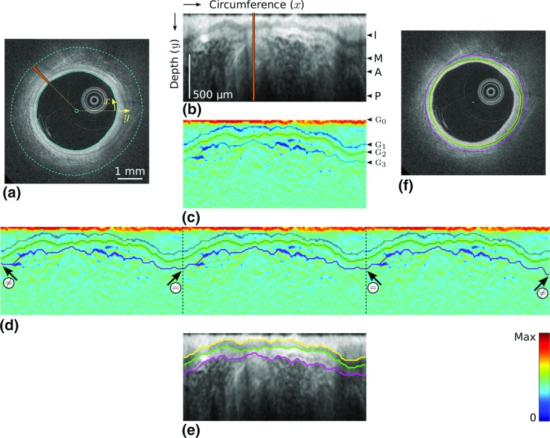
First, the interfaces of the intima, media, and adventitia layers are segmented, by means of an original front propagation scheme, running in a 4D multi-parametric space, to simultaneously extract three non-crossing contours in the initial cross-sectional image. Second, information resulting from the tentative contours is exploited by a machine learning approach to identify healthy and diseased regions of the arterial wall. The framework is fully automatic.
The method was applied to 40 patients from two different medical centers. The framework was trained on 140 images and validated on 260 other images. For the contour segmentation method, the average segmentation errors were [Formula: see text] for the intima-media interface, [Formula: see text] for the media-adventitia interface, and [Formula: see text] for the adventitia-periadventitia interface. The classification method demonstrated a good accuracy, with a median Dice coefficient equal to 0.93 and an interquartile range of (0.78-0.98).
The proposed framework demonstrated promising offline performances and could potentially be translated into a reliable tool for various clinical applications, such as quantification of tissue layer thickness and global summarization of healthy regions in entire pullbacks.
定量和自动分析冠状动脉光学相干断层扫描图像,对于评估临床心血管风险非常有用且节省时间。
首先,通过原始的前向传播方案,在 4D 多参数空间中运行,以同时提取初始横截面图像中的三个非交叉轮廓,从而对内膜、中膜和外膜层的界面进行分割。其次,通过机器学习方法利用来自试探轮廓的信息来识别动脉壁的健康和患病区域。该框架是全自动的。
该方法应用于来自两个不同医疗中心的 40 名患者。该框架在 140 张图像上进行了训练,并在另外 260 张图像上进行了验证。对于轮廓分割方法,平均分割误差分别为[公式:见文本]内膜-中膜界面,[公式:见文本]中膜-外膜界面和[公式:见文本]外膜-外膜周围组织界面。分类方法表现出良好的准确性,中位数 Dice 系数等于 0.93,四分位距为(0.78-0.98)。
所提出的框架表现出有前景的离线性能,并且有可能转化为各种临床应用的可靠工具,例如组织层厚度的定量和整个拉回中健康区域的整体总结。