Department of Neurology, Philipps University Marburg, Baldingerstr, 35033, Marburg, Germany.
Department of Anesthesiology and Intensive Care Medicine, University Hospital, Albert-Schweitzer Campus 1, 48149, Münster, Germany.
Fluids Barriers CNS. 2017 Aug 14;14(1):22. doi: 10.1186/s12987-017-0070-5.
Neoplastic invasion into leptomeninges and subarachnoid space, resulting in neoplastic meningitis (NM) is a fatal complication of advanced solid and hematological neoplasms. Identification of malignant involvement of the cerebrospinal fluid (CSF) early in the disease course has crucial prognostic and therapeutic implications, but remains challenging. As indicators of extracellular matrix (ECM) degradation and breakdown of the blood-brain-barrier, Matrix Metalloproteases (MMPs) and A Disintegrin and Metalloproteases (ADAMs) are potential analytes for cerebral pathophysiology and metastatic dissemination of tumor cells into the CSF.
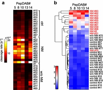
We compared protease activities in CSF samples from patients with NM and control individuals using FRET-based metalloprotease substrates with distinct enzyme selectivity profiles in a real-time, multiplex approach termed "proteolytic activity matrix assay" (PrAMA). Protease activity dynamics can be tracked by fluorescence changes over time. By simultaneously monitoring a panel of 5 FRET-substrate cleavages, a proteolytic signature can be identified and analyzed to infer the activities of multiple specific proteases. Distinct patterns of substrate cleavage comparing disease vs. control samples allow rapid, reproducible and sensitive discrimination even in small volumes of CSF.
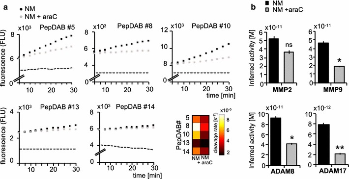
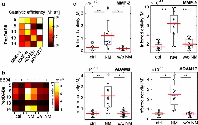
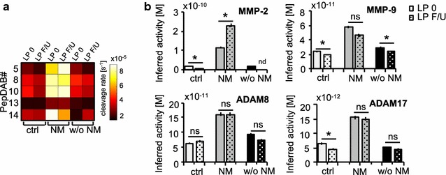
Individual substrate cleavage rates were linked to distinct proteases, and PrAMA computational inference implied increased activities of MMP-9, ADAM8 and ADAM17 (4-5-fold on average) in CSF samples from NM patients that were inhibitable by the metalloprotease inhibitor batimastat (BB-94). The activities of these proteases correlated with blood-brain barrier impairment. Notably, CSF cell counts were not found to directly reflect the protease activities observed in CSF samples from NM patients; this may explain the frequent clinical observation of negative cytology in NM patients.
PrAMA analysis of CSF samples is a potential diagnostic method for sensitive detection of NM and may be suitable for the clinical routine.
肿瘤侵犯软脑膜和蛛网膜下腔,导致癌性脑膜炎(NM),是晚期实体瘤和血液系统恶性肿瘤的致命并发症。在疾病早期识别脑脊液(CSF)中的恶性浸润具有重要的预后和治疗意义,但仍然具有挑战性。作为细胞外基质(ECM)降解和血脑屏障破坏的指标,基质金属蛋白酶(MMPs)和解整合素金属蛋白酶(ADAMs)是脑病理生理学和肿瘤细胞向 CSF 转移的潜在分析物。
我们使用基于荧光共振能量转移(FRET)的基质金属蛋白酶底物,通过实时、多重方法“蛋白水解活性矩阵分析”(PrAMA),比较了 NM 患者和对照个体 CSF 样本中的蛋白酶活性。通过随时间监测荧光变化,可以跟踪蛋白酶活性的动态。通过同时监测 5 个 FRET-底物切割的面板,可以识别和分析蛋白水解特征,以推断多种特定蛋白酶的活性。与对照样本相比,疾病样本的不同底物切割模式允许快速、可重复和敏感的区分,即使在 CSF 体积较小的情况下也是如此。
个体底物切割速率与特定蛋白酶相关,PrAMA 计算推断 MMP-9、ADAM8 和 ADAM17 的活性增加(平均增加 4-5 倍),NM 患者的 CSF 样本中,这些蛋白酶的活性可被金属蛋白酶抑制剂 batimastat(BB-94)抑制。这些蛋白酶的活性与血脑屏障损伤相关。值得注意的是,CSF 细胞计数并未直接反映 NM 患者 CSF 样本中观察到的蛋白酶活性;这可能解释了 NM 患者经常出现细胞学阴性的临床观察。
CSF 样本的 PrAMA 分析是 NM 敏感检测的潜在诊断方法,可能适合临床常规。