Department of obstetrics and gynaecology, Veer Surendra Sai Institute of Medical Science And Research (VIMSAR), Burla, Sambalpur, Odisha, India.
J Ovarian Res. 2017 Aug 14;10(1):55. doi: 10.1186/s13048-017-0351-2.
There is no universal screening method for discrimination between benign and malignant adnexal masses yet. Various authors have tried tumor markers, imaging studies, cytology but no one yet is a definite method for screening of cancer ovary, for which a combined diagnostic modality has come to practice in form of RMI. With this background we conducted our study "Evaluation of risk malignancy index and its diagnostic value in patients with adnexal masses".
The aim of the study was to determine the effectiveness of risk of malignancy index (RMI-3) in preoperative discrimination between benign and malignant masses and also to reveal the most suitable cut off value. We have conducted a prospective study between November 2014 to October 2016. We included the parameters like menopausal status, ultrasound features, and serum levels of tumor marker like CA-125 for calculating RMI 3. Then RMI was compared with the histopathological report which was taken as gold standard.
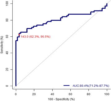
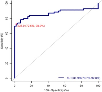
In the present study malignant tumors constitute 54.76% (69/126) & benign tumors 45.24% (57/126). Bilaterality in adnexal masses and multilocularity is higher in malignant tumors than benign tumor, but a P -value >0.005 failed to be proved significant in our study. Solid area is seen in 24.69% (20/81) of benign and 75.30% (61/81) of malignant tumor. Similarly ascites was found in 38.09% (48/126) of cases. Out of which 18.75% (9/48) cases were found to be benign and malignancy was confirmed in 81.25% (39/48) patients. There is statistically significant number of malignant ovarian cancer patients where ascites and solid area is seen in USG findings (p = 0.000). Risk of Malignancy Index compared with individual parameters of Ultrasound score, CA-125 or menopausal score and a cut-off point of 236 shows a very high sensitivity (72.5%), specificity (98.2%), positive predictive value (98.1%), negative predictive value (74.7%) and diagnostic accuracy (84.13%) for discriminating malignant and benign pelvic masses.
Simplicity and applicability of the method in the primary evaluation of patients with pelvic masses makes it a good option in daily clinical practice in non-specialized gynecologic departments and also in developing countries where access to a gynaecologist oncologist is limited.
目前尚无普遍适用于鉴别附件区良恶性包块的方法。许多作者尝试了肿瘤标志物、影像学研究和细胞学检查,但尚无一种方法可明确筛查卵巢癌,目前已采用 RMI 联合诊断模式。基于此背景,我们开展了“评价风险恶性指数及其在附件区包块患者中的诊断价值”的研究。
本研究旨在确定术前风险恶性指数(RMI-3)在鉴别良恶性包块中的有效性,并揭示最适宜的截断值。我们于 2014 年 11 月至 2016 年 10 月开展了一项前瞻性研究。纳入绝经状态、超声特征和血清肿瘤标志物 CA-125 等参数来计算 RMI-3。然后,将其与作为金标准的组织病理学报告进行比较。
在本研究中,恶性肿瘤占 54.76%(69/126),良性肿瘤占 45.24%(57/126)。附件区包块的双侧性和多房性在恶性肿瘤中比良性肿瘤更为常见,但 P 值>0.005 未能证明其具有显著差异。良性肿瘤中 24.69%(20/81)有实性区,而恶性肿瘤中 75.30%(61/81)有实性区。同样,腹水见于 126 例中的 38.09%(48/126)。其中 18.75%(9/48)为良性,81.25%(39/48)为恶性肿瘤。在超声检查结果中,恶性卵巢癌患者中腹水和实性区的数量有统计学意义(p=0.000)。与超声评分、CA-125 或绝经评分的个别参数相比,风险恶性指数和 236 的截断值显示出非常高的灵敏度(72.5%)、特异性(98.2%)、阳性预测值(98.1%)、阴性预测值(74.7%)和诊断准确性(84.13%),可用于鉴别盆腔良恶性包块。
该方法在盆腔包块患者的初步评估中具有简单易用的特点,使其成为非专科妇科和卵巢癌专科医生资源有限的发展中国家的常规临床实践中的良好选择。