Hwang Do Won, Kwon Hyun Woo, Jang Jaeho, Jung Hee Jung, Kim Kwang Rok, Lee Dong Soo
Department of Nuclear Medicine, Seoul National University College of Medicine, Seoul, Republic of Korea.
Department of Molecular Medicine and Biopharmaceutical Sciences, Graduate School of Convergence Science and Technology and College of Medicine or College of Pharmacy, Seoul National University, Seoul, Republic of Korea.
Stem Cells Int. 2017;2017:8452830. doi: 10.1155/2017/8452830. Epub 2017 Jul 20.
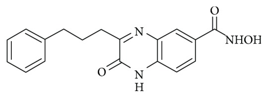
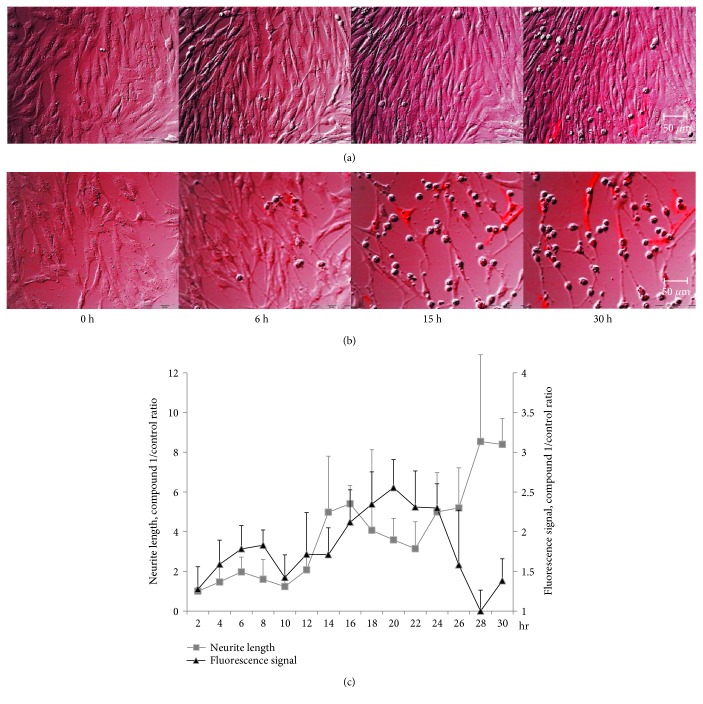
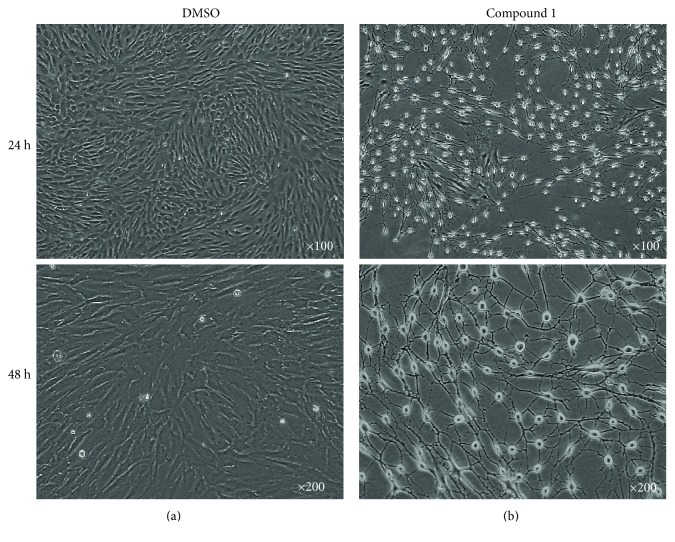
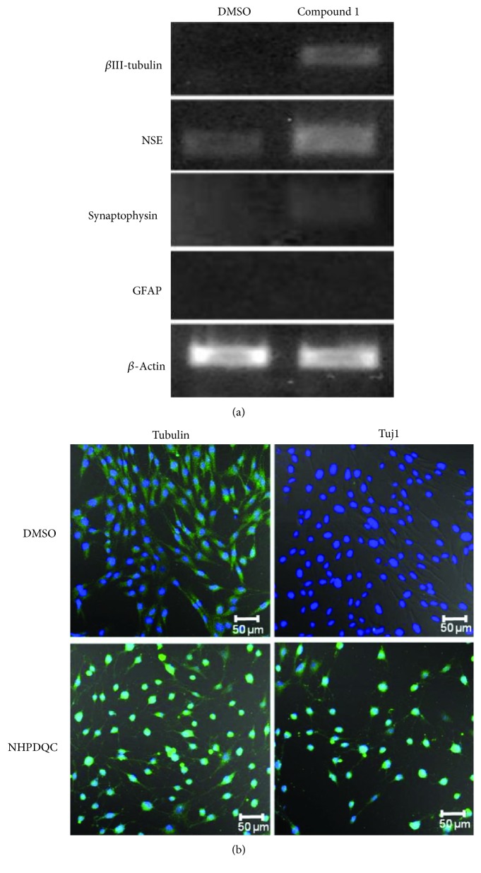
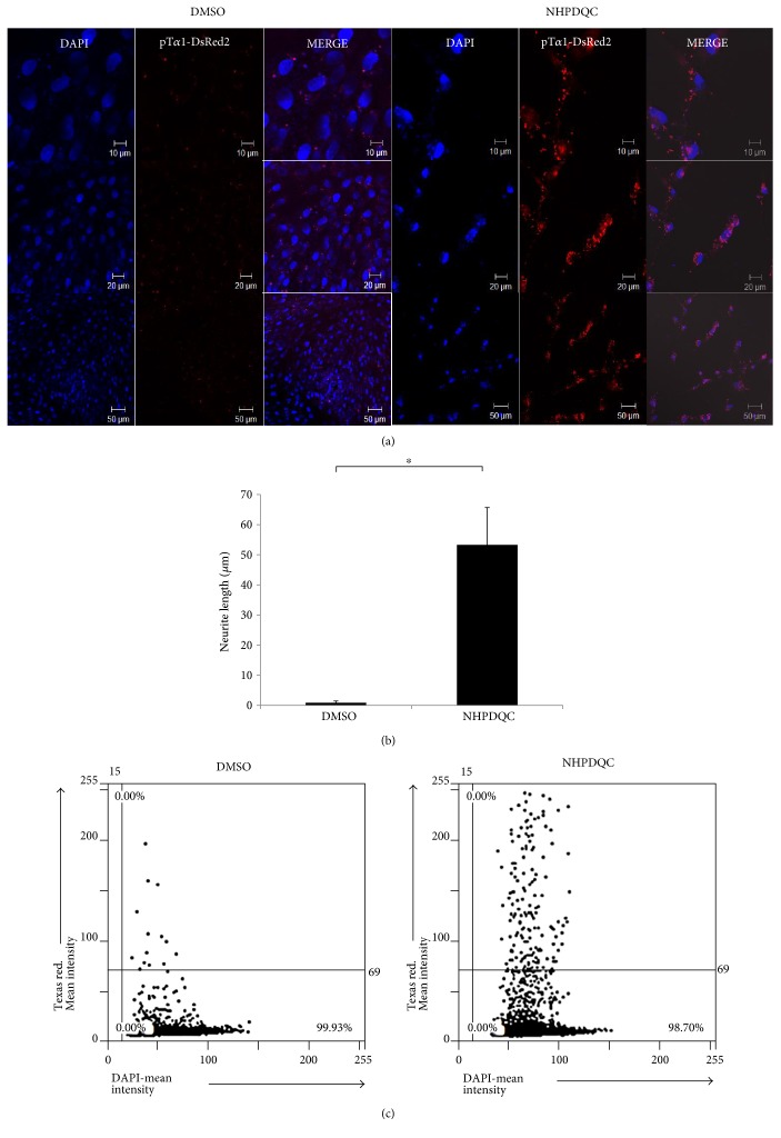
Although transdifferentiation of mesenchymal stem cells (MSCs) into neurons increases the possibility of therapeutic use of MSCs for neurodevelopmental disorders, the use of MSCs has the limitation on differentiation efficiency to neuronal lineage and lack of an easy method to monitor the transdifferentiation. In this study, using time-lapse live cell imaging, we assessed the neuronal differentiation of MSCs induced by a small molecule "NHPDQC (N-hydroxy-2-oxo-3-(3-phenylprophyl)-1,2-dihydroquinoxaline-6-carboxamide, CHNO)." Plasmid vector containing red fluorescence reporter genes under the control of the tubulin 1 (T1) promoter (pT1-DsRed2) traced the neuronal differentiation of MSCs. Two days after NHPDQC treatment, MSCs showed neuron-like phenotype with neurite outgrowth and high expression of neuron-specific markers in more than 95% cells. The fluorescence signals increased in the cytoplasm of pT1-DsRed2-transfected MSCs after NHPDQC treatment. In vitro monitoring of MSCs along the time courses showed progressive increase of fluorescence till 30 h after treatment, corresponding with the increase in neurite length. We examined an efficient neuronal differentiation of MSCs by NHPDQC alone and monitored the temporal changes of neuronal differentiation by neuron-specific fluorescence reporter along time. This method would help further our understanding of the differentiation of MSCs to produce neurons by simple treatment of small molecule.
尽管间充质干细胞(MSCs)向神经元的转分化增加了将MSCs用于神经发育障碍治疗的可能性,但MSCs的使用在向神经谱系的分化效率方面存在局限性,并且缺乏监测转分化的简便方法。在本研究中,我们使用延时活细胞成像技术,评估了小分子“NHPDQC(N-羟基-2-氧代-3-(3-苯基丙基)-1,2-二氢喹喔啉-6-甲酰胺,CHNO)”诱导的MSCs的神经元分化。含有在微管蛋白1(T1)启动子控制下的红色荧光报告基因的质粒载体(pT1-DsRed2)追踪了MSCs的神经元分化。NHPDQC处理两天后,MSCs呈现出神经元样表型,有神经突生长,并且在超过95%的细胞中高表达神经元特异性标志物。NHPDQC处理后,pT1-DsRed2转染的MSCs细胞质中的荧光信号增强。对MSCs进行的体外时间进程监测显示,荧光在处理后30小时内逐渐增加,这与神经突长度的增加相对应。我们研究了单独使用NHPDQC时MSCs的高效神经元分化情况,并通过神经元特异性荧光报告基因随时间监测神经元分化的动态变化。这种方法将有助于我们进一步了解通过简单的小分子处理使MSCs分化产生神经元的过程。