Columbia University Department of Otolaryngology/Head and Neck Surgery, United States.
Columbia University Department of Radiology, United States.
Neuroimage Clin. 2017 Jul 24;16:205-209. doi: 10.1016/j.nicl.2017.07.021. eCollection 2017.
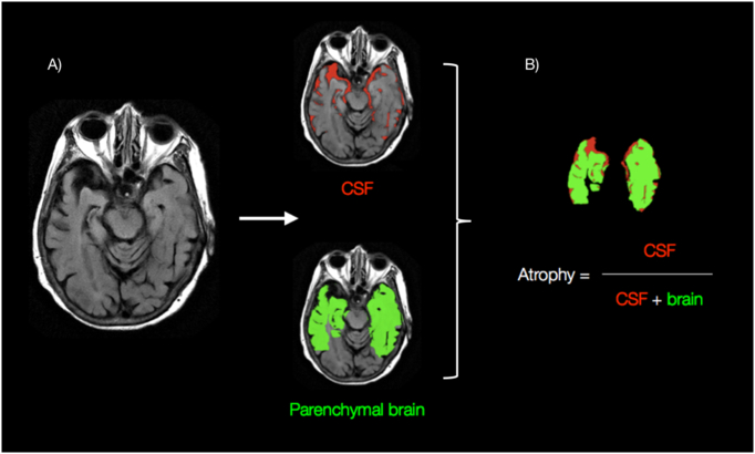
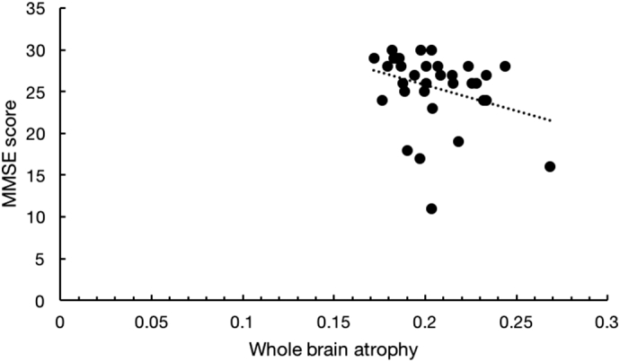
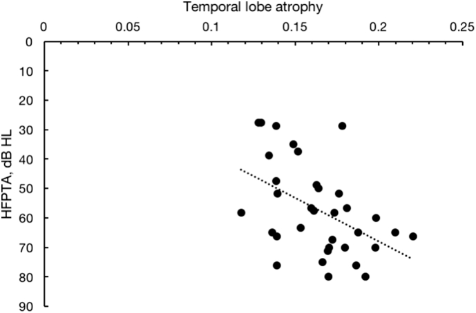
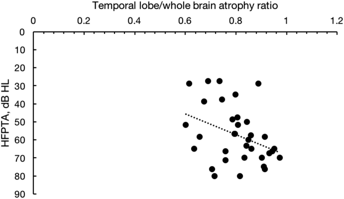
A growing body of evidence has shown that a relationship between age-related hearing loss and structural brain changes exists. However, a method to measure brain atrophy associated with hearing loss from a single MRI study (i.e. without an interval study) that produces an independently interpretable output does not. Such a method would be beneficial for studying patterns of structural brain changes on a large scale. Here, we introduce our method for this. Audiometric evaluations and mini-mental state exams were obtained in 34 subjects over the age of 80 who have had brain MRIs in the past 6 years. CSF and parenchymal brain volumes (whole brain and by lobe) were obtained through a novel, fully automated algorithm. Atrophy was calculated by taking the ratio of CSF to parenchyma. High frequency hearing loss was associated with disproportional temporal lobe atrophy relative to whole brain atrophy independent of age ( = 0.471, p = 0.005). Mental state was associated with frontoparietal atrophy but not to temporal lobe atrophy, which is consistent with known results. Our method demonstrates that hearing loss is associated with temporal lobe atrophy and generalized whole brain atrophy. Our algorithm is efficient, fully automated, and able to detect significant associations in a small cohort.
越来越多的证据表明,与年龄相关的听力损失与结构性脑变化之间存在关系。然而,目前还没有一种方法可以从单次 MRI 研究(即没有间隔研究)中测量与听力损失相关的脑萎缩,并产生独立可解释的结果。这种方法将有利于大规模研究结构脑变化的模式。在这里,我们介绍了这种方法。对过去 6 年内接受过脑部 MRI 的 34 名 80 岁以上的受试者进行了听力评估和简易精神状态检查。通过一种新颖的全自动算法获得 CSF 和脑实质体积(全脑和各叶)。通过 CSF 与脑实质的比值计算萎缩程度。高频听力损失与全脑萎缩相比,与颞叶不成比例的萎缩相关,与年龄无关(= 0.471,p = 0.005)。精神状态与额顶叶萎缩有关,但与颞叶萎缩无关,这与已知的结果一致。我们的方法表明听力损失与颞叶萎缩和全脑普遍萎缩有关。我们的算法效率高、完全自动化,可以在小队列中检测到显著的相关性。