Helliwell Jack A, Thomas Daniel S, Papathanasiou Vaia, Homer-Vanniasinkam Shervanthi, Desai Amisha, Jennings Louise M, Rooney Paul, Kearney John N, Ingham Eileen
Institute of Medical and Biological Engineering, University of Leeds, Leeds, UK.
Leeds Vascular Institute, Leeds General Infirmary, Leeds, UK.
J Tissue Eng. 2017 Aug 3;8:2041731417724011. doi: 10.1177/2041731417724011. eCollection 2017 Jan-Dec.
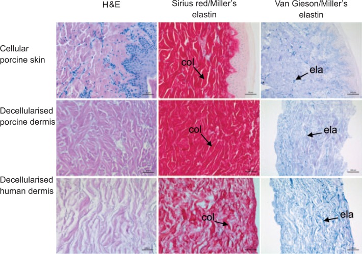
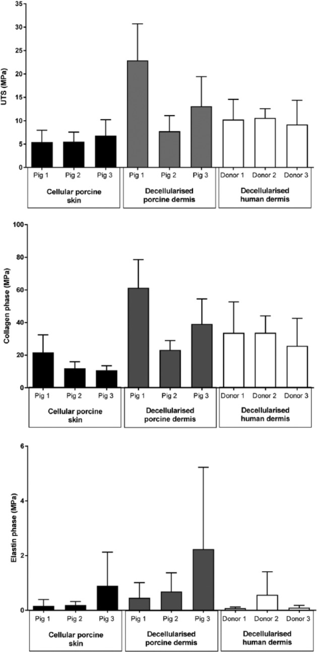
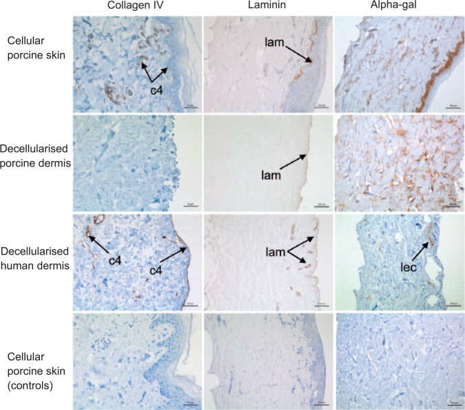
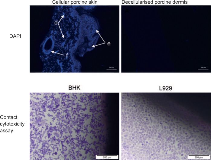
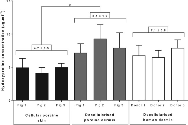
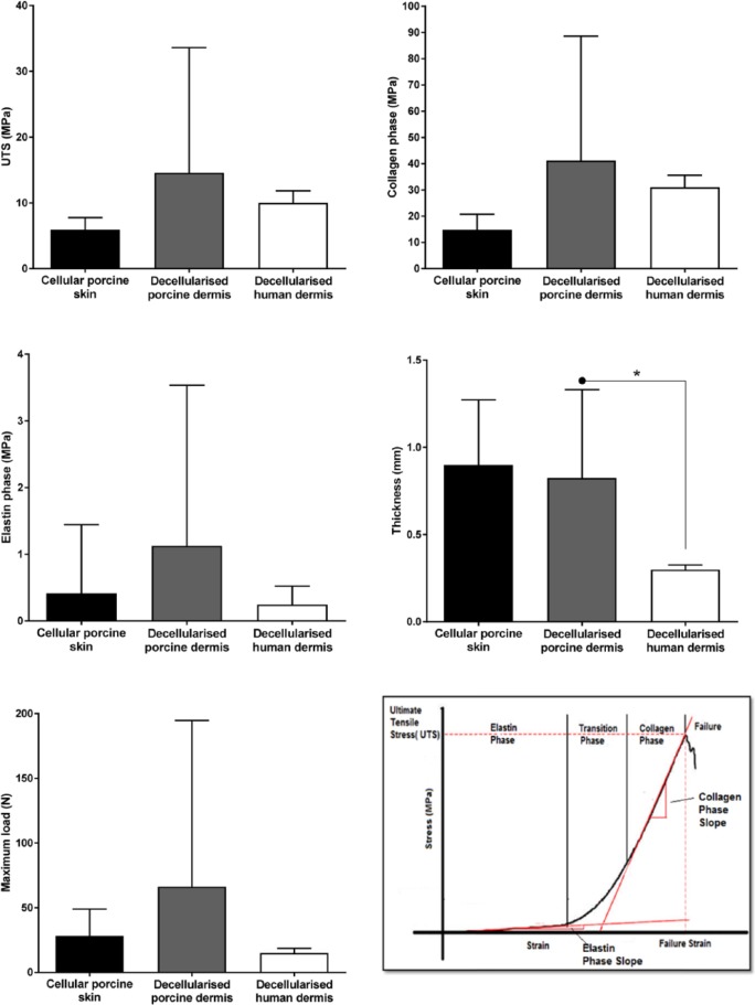
The aim of this study was to adapt a proprietary decellularisation process for human dermis for use with porcine skin. Porcine skin was subject to: sodium chloride (1 M) to detach the epidermis, trypsin paste to remove hair follicles, peracetic acid (0.1% v/v) disinfection, washed in hypotonic buffer and 0.1% (w/v) sodium dodecyl sulphate in the presence of proteinase inhibitors followed by nuclease treatment. Cellular porcine skin, decellularised porcine and human dermis were compared using histology, immunohistochemistry, GSL-1 lectin (alpha-gal epitope) staining, biochemical assays, uniaxial tensile and in vitro cytotoxicity tests. There was no microscopic evidence of cells in decellularised porcine dermis. DNA content was reduced by 98.2% compared to cellular porcine skin. There were no significant differences in the biomechanical parameters studied or evidence of cytotoxicity. The decellularised porcine dermis retained residual alpha-gal epitope. Basement membrane collagen IV immunostaining was lost following decellularisation; however, laminin staining was retained.
本研究的目的是调整一种用于人类真皮的专有去细胞化方法,使其适用于猪皮。猪皮经过以下处理:用氯化钠(1M)分离表皮,用胰蛋白酶糊剂去除毛囊,用过氧乙酸(0.1% v/v)消毒,在低渗缓冲液中洗涤,然后在蛋白酶抑制剂存在的情况下用0.1%(w/v)十二烷基硫酸钠处理,随后进行核酸酶处理。使用组织学、免疫组织化学、GSL-1凝集素(α-半乳糖表位)染色、生化分析、单轴拉伸和体外细胞毒性试验对细胞性猪皮、去细胞化猪皮和人类真皮进行了比较。在去细胞化猪真皮中没有细胞的微观证据。与细胞性猪皮相比,DNA含量降低了98.2%。所研究的生物力学参数没有显著差异,也没有细胞毒性的证据。去细胞化猪真皮保留了残留的α-半乳糖表位。去细胞化后,基底膜胶原蛋白IV免疫染色消失;然而,层粘连蛋白染色得以保留。