Department of Surgery, University of Massachusetts Medical School, Worcester, MA 01655, USA.
J Immunol Res. 2015;2015:589648. doi: 10.1155/2015/589648. Epub 2015 Apr 1.
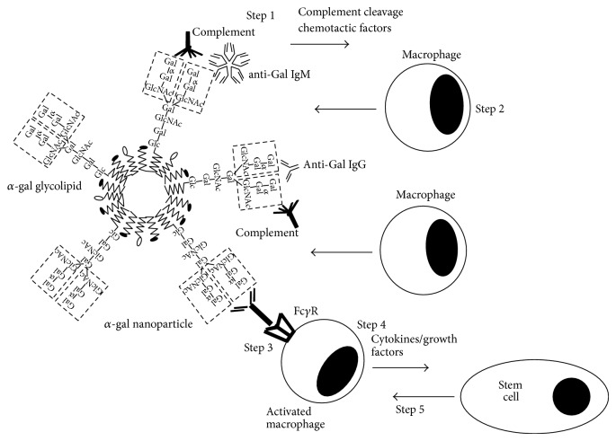
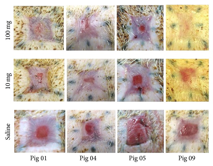
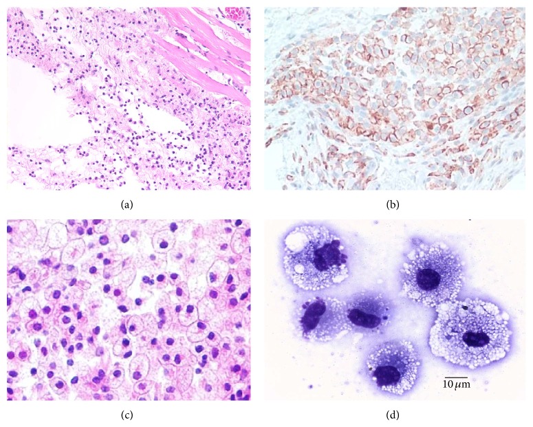
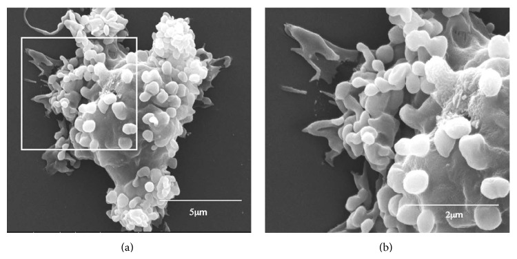
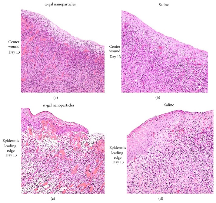
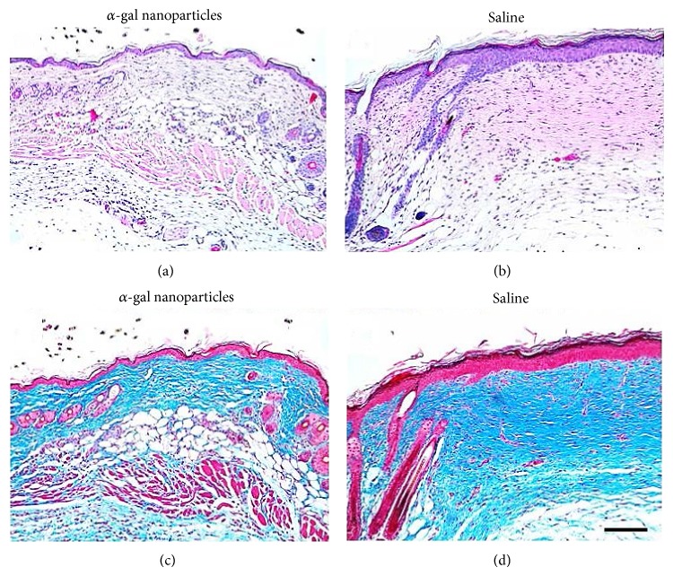
Application of α-gal nanoparticles to wounds and burns induces accelerated healing by harnessing the natural anti-Gal antibody which constitutes ~1% of human immunoglobulins. α-gal nanoparticles present multiple α-gal epitopes (Galα1-3Galβ1-4GlcNAc-R), the carbohydrate ligand of anti-Gal. Studied α-gal nanoparticles were comprised of glycolipids with α-gal epitopes, phospholipids, and cholesterol. Binding of anti-Gal to α-gal nanoparticles in wounds activates the complement cascade, resulting in formation of chemotactic complement cleavage peptides that induce rapid recruitment of many macrophages. The Fc/Fcγ receptors interaction between anti-Gal coating α-gal nanoparticles and the recruited macrophages activates macrophages to produce cytokines/growth factors that promote wound healing and recruit stem cells. Studies of wound healing by α-gal nanoparticles were feasible in α1,3galactosyltransferase knockout mice and pigs. In contrast to other nonprimate mammals, these mice and pigs lack the α-gal epitope, and thus they are not immunotolerant to it and produce anti-Gal. Treatment of skin wounds and burns with α-gal nanoparticles resulted in 40-60% decrease in healing time in comparison with control wounds treated with saline. This accelerated healing is associated with increased recruitment of macrophages and extensive angiogenesis in wounds, faster regrowth of epidermis, and regeneration of the dermis. The accelerated healing further decreases and may completely eliminate fibrosis and scar formation in wounds. Since healing of internal injuries is mediated by mechanisms similar to those in external wound healing, it is suggested that α-gal nanoparticles treatment may also improve regeneration and restoration of biological function following internal injuries such as surgical incisions, myocardial ischemia following infarction, and nerve injuries.
α-半乳糖纳米颗粒在伤口和烧伤中的应用通过利用构成人类免疫球蛋白约 1%的天然抗-Gal 抗体来加速愈合。α-半乳糖纳米颗粒呈现多个α-半乳糖表位(Galα1-3Galβ1-4GlcNAc-R),即抗-Gal 的碳水化合物配体。研究中使用的α-半乳糖纳米颗粒由带α-半乳糖表位的糖脂、磷脂和胆固醇组成。抗-Gal 与伤口中的α-半乳糖纳米颗粒结合会激活补体级联反应,导致趋化性补体裂解肽的形成,从而迅速募集大量巨噬细胞。抗-Gal 涂层α-半乳糖纳米颗粒与募集的巨噬细胞之间的 Fc/Fcγ 受体相互作用激活巨噬细胞产生细胞因子/生长因子,促进伤口愈合并募集干细胞。α-半乳糖纳米颗粒在α1,3-半乳糖基转移酶敲除小鼠和猪中的伤口愈合研究是可行的。与其他非灵长类哺乳动物不同,这些小鼠和猪缺乏α-半乳糖表位,因此它们对其没有免疫耐受性,并且会产生抗-Gal。与用生理盐水处理的对照伤口相比,用α-半乳糖纳米颗粒治疗皮肤伤口和烧伤可使愈合时间缩短 40-60%。这种加速愈合与巨噬细胞的大量募集和伤口中广泛的血管生成、表皮更快的再生以及真皮的再生有关。加速愈合进一步减少甚至可能完全消除伤口中的纤维化和瘢痕形成。由于内部损伤的愈合是通过与外部伤口愈合相似的机制介导的,因此建议α-半乳糖纳米颗粒治疗也可能改善手术后切口、梗塞后心肌缺血和神经损伤等内部损伤后的再生和恢复生物功能。