• 文献检索
  • 文档翻译
  • 深度研究
  • 学术资讯
  • Suppr Zotero 插件Zotero 插件
  • 邀请有礼
  • 套餐&价格
  • 历史记录
应用&插件
Suppr Zotero 插件Zotero 插件浏览器插件Mac 客户端Windows 客户端微信小程序
定价
高级版会员购买积分包购买API积分包
服务
文献检索文档翻译深度研究API 文档MCP 服务
关于我们
关于 Suppr公司介绍联系我们用户协议隐私条款
关注我们

Suppr 超能文献

核心技术专利:CN118964589B侵权必究
粤ICP备2023148730 号-1Suppr @ 2026

文献检索

告别复杂PubMed语法,用中文像聊天一样搜索,搜遍4000万医学文献。AI智能推荐,让科研检索更轻松。

立即免费搜索

文件翻译

保留排版,准确专业,支持PDF/Word/PPT等文件格式,支持 12+语言互译。

免费翻译文档

深度研究

AI帮你快速写综述,25分钟生成高质量综述,智能提取关键信息,辅助科研写作。

立即免费体验

活体多光子显微镜在小鼠耳蜗可视化研究及中阶容积变化研究中的应用价值

Usefulness of Intravital Multiphoton Microscopy in Visualizing Study of Mouse Cochlea and Volume Changes in the Scala Media.

作者信息

Ju Hyun Mi, Lee Sun Hee, Kong Tae Hoon, Kwon Seung-Hae, Choi Jin Sil, Seo Young Joon

机构信息

Laboratory of Smile Snail, Yonsei University Wonju College of Medicine, Wonju, South Korea.

Department of Otorhinolaryngology, Yonsei University Wonju College of Medicine, Wonju, South Korea.

出版信息

Front Neurol. 2017 Jul 31;8:332. doi: 10.3389/fneur.2017.00332. eCollection 2017.

DOI:10.3389/fneur.2017.00332
PMID:28824523
原文链接:https://pmc.ncbi.nlm.nih.gov/articles/PMC5535263/
Abstract

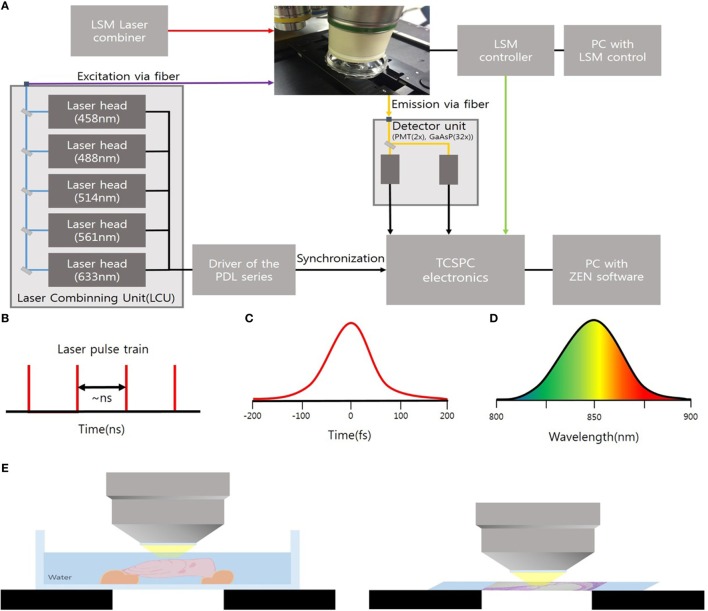
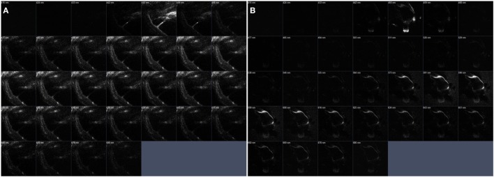
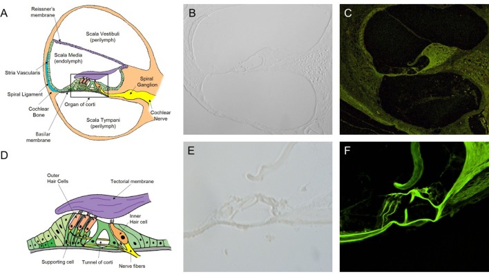
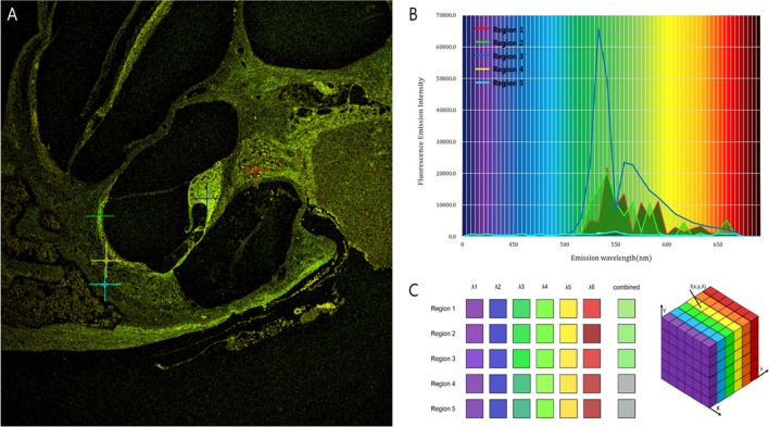
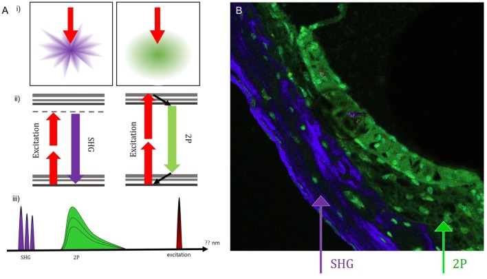
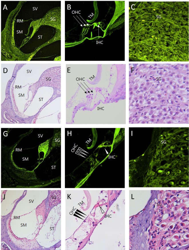
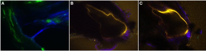
Conventional microscopy has limitations in viewing the cochlear microstructures due to three-dimensional spiral structure and the overlying bone. But these issues can be overcome by imaging the cochlea with intravital multiphoton microscopy (MPM). By using near-infrared lasers for multiphoton excitation, intravital MPM can detect endogenous fluorescence and second harmonic generation of tissues. In this study, we used intravital MPM to visualize various cochlear microstructures without any staining and non-invasively analyze the volume changes of the scala media (SM) without removing the overlying cochlear bone. The intravital MPM images revealed various tissue types, ranging from thin membranes to dense bone, as well as the spiral ganglion beneath the cochlear bone. The two-dimensional, cross-sectional, and serial z-stack intravital MPM images also revealed the spatial dilation of the SM in the temporal bone of pendrin-deficient mice. These findings suggest that intravital MPM might serve as a new method for obtaining microanatomical information regarding the cochlea, similar to standard histopathological analyses in the animal study for the cochlea. Given the capability of intravital MPM for detecting an increase in the volume of the SM in pendrin-deficient mice, it might be a promising new tool for assessing the pathophysiology of hearing loss in the future.

摘要

由于三维螺旋结构和覆盖其上的骨骼,传统显微镜在观察耳蜗微观结构方面存在局限性。但通过活体多光子显微镜(MPM)对耳蜗进行成像,可以克服这些问题。通过使用近红外激光进行多光子激发,活体MPM能够检测组织的内源性荧光和二次谐波产生。在本研究中,我们使用活体MPM在不进行任何染色的情况下可视化各种耳蜗微观结构,并在不去除覆盖的耳蜗骨的情况下非侵入性地分析中阶(SM)的体积变化。活体MPM图像显示了从薄膜到致密骨骼等各种组织类型,以及耳蜗骨下方的螺旋神经节。二维、横截面和连续z轴堆叠的活体MPM图像还显示了pendrin缺陷小鼠颞骨中SM的空间扩张。这些发现表明,活体MPM可能成为获取有关耳蜗微观解剖信息的一种新方法,类似于动物耳蜗研究中的标准组织病理学分析。鉴于活体MPM能够检测pendrin缺陷小鼠中SM体积的增加,它可能是未来评估听力损失病理生理学的一种有前景的新工具。

https://cdn.ncbi.nlm.nih.gov/pmc/blobs/615e/5535263/8931918e0886/fneur-08-00332-g007.jpg
https://cdn.ncbi.nlm.nih.gov/pmc/blobs/615e/5535263/dd624a1ee913/fneur-08-00332-g001.jpg
https://cdn.ncbi.nlm.nih.gov/pmc/blobs/615e/5535263/9b7f8c76ef5c/fneur-08-00332-g002.jpg
https://cdn.ncbi.nlm.nih.gov/pmc/blobs/615e/5535263/98fd69e11db0/fneur-08-00332-g003.jpg
https://cdn.ncbi.nlm.nih.gov/pmc/blobs/615e/5535263/3e9a78dd66f3/fneur-08-00332-g004.jpg
https://cdn.ncbi.nlm.nih.gov/pmc/blobs/615e/5535263/cc0bf14401d0/fneur-08-00332-g005.jpg
https://cdn.ncbi.nlm.nih.gov/pmc/blobs/615e/5535263/e17da02cd222/fneur-08-00332-g006.jpg
https://cdn.ncbi.nlm.nih.gov/pmc/blobs/615e/5535263/8931918e0886/fneur-08-00332-g007.jpg

相似文献

1
Usefulness of Intravital Multiphoton Microscopy in Visualizing Study of Mouse Cochlea and Volume Changes in the Scala Media.活体多光子显微镜在小鼠耳蜗可视化研究及中阶容积变化研究中的应用价值
Front Neurol. 2017 Jul 31;8:332. doi: 10.3389/fneur.2017.00332. eCollection 2017.
2
Correlation between word recognition score and intracochlear new bone and fibrous tissue after cochlear implantation in the human.人工耳蜗植入术后人类单词识别分数与耳蜗内新骨及纤维组织之间的相关性
Hear Res. 2016 Sep;339:132-41. doi: 10.1016/j.heares.2016.06.015. Epub 2016 Jun 29.
3
Intravital Multiphoton Imaging of the Kidney: Tubular Structure and Metabolism.肾脏的活体多光子成像:肾小管结构与代谢
Methods Mol Biol. 2016;1397:155-172. doi: 10.1007/978-1-4939-3353-2_12.
4
Frontiers in Intravital Multiphoton Microscopy of Cancer.肿瘤活体多光子显微镜前沿
Cancer Rep (Hoboken). 2020 Feb;3(1):e1192. doi: 10.1002/cnr2.1192. Epub 2019 Jun 20.
5
Quantitative X-ray tomography of the mouse cochlea.小鼠耳蜗的定量 X 射线断层扫描。
PLoS One. 2012;7(4):e33568. doi: 10.1371/journal.pone.0033568. Epub 2012 Apr 2.
6
Visualize and quantify the structural alteration of the rat spinal cord injury based on multiphoton microscopy.基于多光子显微镜可视化并量化大鼠脊髓损伤的结构改变。
Lasers Med Sci. 2019 Apr;34(3):561-569. doi: 10.1007/s10103-018-2630-6. Epub 2018 Sep 9.
7
Novel in vivo techniques to visualize kidney anatomy and function.用于可视化肾脏解剖结构和功能的新型体内技术。
Kidney Int. 2015 Jul;88(1):44-51. doi: 10.1038/ki.2015.65. Epub 2015 Mar 4.
8
Cochlear pathology following reimplantation of a multichannel scala tympani electrode array in the macaque.猕猴中多通道鼓阶电极阵列再植入后的耳蜗病理学
Am J Otol. 1995 Mar;16(2):186-99.
9
Recent advances in intravital imaging of dynamic biological systems.动态生物系统活体成像的最新进展。
J Pharmacol Sci. 2012;119(3):193-7. doi: 10.1254/jphs.12r03cp. Epub 2012 Jun 13.
10
Development of the mouse cochlea database (MCD).小鼠耳蜗数据库(MCD)的开发。
Hear Res. 2008 Sep;243(1-2):11-7. doi: 10.1016/j.heares.2008.04.014. Epub 2008 May 25.

引用本文的文献

1
Protection of Hearing Loss in Ototoxic Mouse Model Through SPIONs and Dexamethasone-Loaded PLGA Nanoparticle Delivery by Magnetic Attraction.通过磁吸引递送 SPIONs 和载地塞米松的 PLGA 纳米颗粒保护耳毒性小鼠模型中的听力损失。
Int J Nanomedicine. 2022 Dec 13;17:6317-6334. doi: 10.2147/IJN.S380810. eCollection 2022.
2
Neutrophils infiltrate into the spiral ligament but not the stria vascularis in the cochlea during lipopolysaccharide-induced inflammation.中性粒细胞在脂多糖诱导的炎症期间浸润到螺旋韧带,但未浸润到耳蜗中的血管纹。
Theranostics. 2021 Jan 1;11(6):2522-2533. doi: 10.7150/thno.49121. eCollection 2021.
3

本文引用的文献

1
Experimental Fusion of Contrast Enhanced High-Field Magnetic Resonance Imaging and High-Resolution Micro-Computed Tomography in Imaging the Mouse Inner Ear.对比增强高场磁共振成像与高分辨率微型计算机断层扫描在小鼠内耳成像中的实验融合
Open Neuroimag J. 2015 Jul 31;9:7-12. doi: 10.2174/1874440001509010007. eCollection 2015.
2
Two-photon microscopy allows imaging and characterization of cochlear microvasculature in vivo.双光子显微镜能够对体内耳蜗微血管进行成像和特征分析。
Biomed Res Int. 2015;2015:154272. doi: 10.1155/2015/154272. Epub 2015 Mar 30.
3
Micro CT visualization of silver nanoparticles in the middle and inner ear of rat and transportation pathway after transtympanic injection.
Label-Free Visualization and Tracking of Gold Nanoparticles in Vasculature Using Multiphoton Luminescence.
利用多光子发光对血管中的金纳米颗粒进行无标记可视化和追踪
Nanomaterials (Basel). 2020 Nov 12;10(11):2239. doi: 10.3390/nano10112239.
4
Chronic cigarette smoke exposure drives spiral ganglion neuron loss in mice.慢性香烟烟雾暴露导致小鼠耳蜗螺旋神经节神经元丧失。
Sci Rep. 2018 Apr 10;8(1):5746. doi: 10.1038/s41598-018-24166-9.
5
A Simple Model for Inducing Optimal Increase of with Aminoglycoside Ototoxicity.一种用于诱导氨基糖苷耳毒性中 最佳增加的简单模型。
Biomed Res Int. 2017;2017:4630241. doi: 10.1155/2017/4630241. Epub 2017 Dec 21.
大鼠中耳和内耳银纳米颗粒的显微CT可视化及经鼓膜注射后的转运途径
J Nanobiotechnology. 2015 Jan 27;13:5. doi: 10.1186/s12951-015-0065-9.
4
Developmental changes of ENaC expression and function in the inner ear of pendrin knock-out mice as a perspective on the development of endolymphatic hydrops.作为对内淋巴积水发展的一种展望,探讨pendrin基因敲除小鼠内耳中ENaC表达和功能的发育变化。
PLoS One. 2014 Apr 21;9(4):e95730. doi: 10.1371/journal.pone.0095730. eCollection 2014.
5
Multiphoton fluorescence microscopy of the live kidney in health and disease.活体肾脏的多光子荧光显微镜检查:健康与疾病。
J Biomed Opt. 2014 Feb;19(2):020901. doi: 10.1117/1.JBO.19.2.020901.
6
Kanamycin-furosemide ototoxicity in the mouse cochlea: a 3-dimensional analysis.卡那霉素-呋塞米耳毒性在小鼠耳蜗中的三维分析。
Otolaryngol Head Neck Surg. 2014 Apr;150(4):666-72. doi: 10.1177/0194599813519071. Epub 2014 Jan 10.
7
Advances in multiphoton microscopy technology.多光子显微镜技术的进展。
Nat Photonics. 2013 Feb 1;7(2):93-101. doi: 10.1038/nphoton.2012.361.
8
High-resolution three-photon biomedical imaging using doped ZnS nanocrystals.使用掺杂 ZnS 纳米晶体的高分辨率三光子生物医学成像。
Nat Mater. 2013 Apr;12(4):359-66. doi: 10.1038/nmat3565. Epub 2013 Feb 17.
9
Visualization of endolymphatic hydrops and correlation with audio-vestibular functional testing in patients with definite Meniere's disease.确诊梅尼埃病患者内淋巴积水的可视化及其与听-前庭功能测试的相关性。
Auris Nasus Larynx. 2013 Apr;40(2):167-72. doi: 10.1016/j.anl.2012.07.009. Epub 2012 Aug 4.
10
Multiphoton microscopy and microspectroscopy for diagnostics of inflammatory and neoplastic lung.多光子显微镜和微光谱学在炎症和肿瘤性肺疾病诊断中的应用。
J Biomed Opt. 2012 Mar;17(3):036014. doi: 10.1117/1.JBO.17.3.036014.