Henao Agudelo Juan S, Braga Tarcio T, Amano Mariane T, Cenedeze Marcos A, Cavinato Regiane A, Peixoto-Santos Amandda R, Muscará Marcelo N, Teixeira Simone A, Cruz Mario C, Castoldi Angela, Sinigaglia-Coimbra Rita, Pacheco-Silva Alvaro, de Almeida Danilo C, Camara Niels Olsen Saraiva
Department of Medicine, Division of Nephrology, Federal University of São Paulo, Sao Paulo, Brazil.
Department of Immunology, Institute of Biomedical Sciences, University of São Paulo, Sao Paulo, Brazil.
Front Immunol. 2017 Jul 31;8:881. doi: 10.3389/fimmu.2017.00881. eCollection 2017.
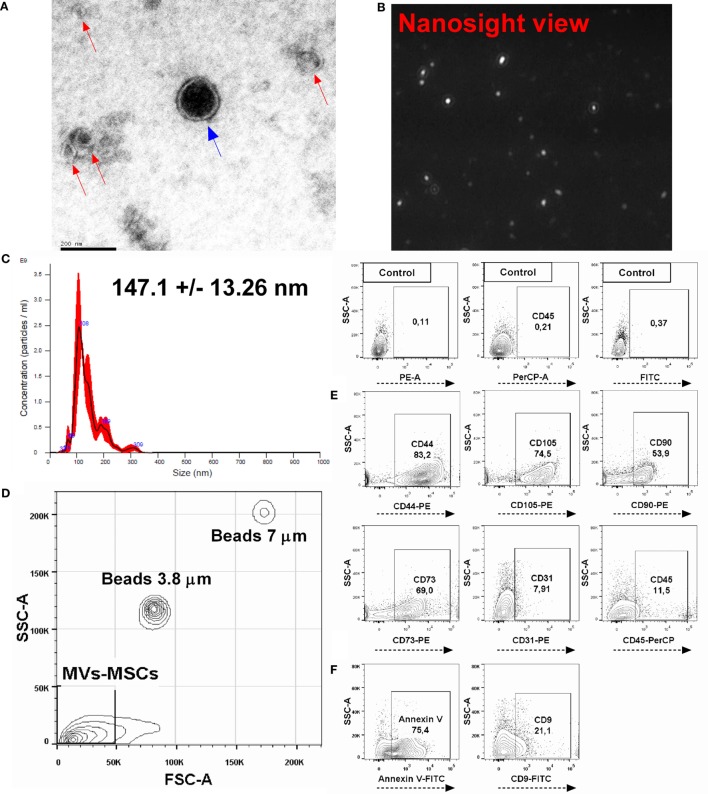
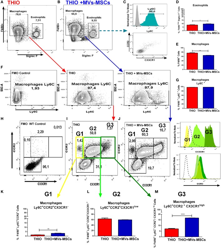
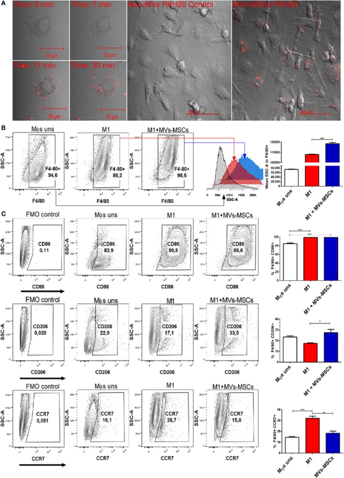
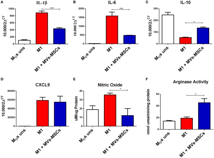
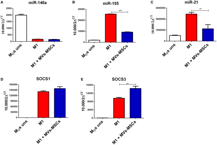
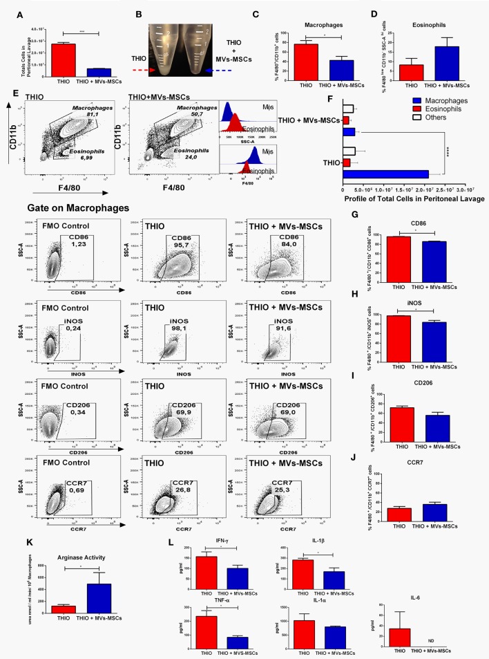
Mesenchymal stromal cells (MSCs) are multipotent cells with abilities to exert immunosuppressive response promoting tissue repair. Studies have shown that MSCs can secrete extracellular vesicles (MVs-MSCs) with similar regulatory functions to the parental cells. Furthermore, strong evidence suggesting that MVs-MSCs can modulate several immune cells (i.e., Th1, Th17, and Foxp3 T cells). However, their precise effect on macrophages (Mϕs) remains unexplored. We investigated the immunoregulatory effect of MVs-MSCs on activated M1-Mϕs and using differentiated bone marrow Mϕs and an acute experimental model of thioglycollate-induced peritonitis, respectively. We observed that MVs-MSCs shared surface molecules with MSCs (CD44, CD105, CD90, CD73) and expressed classical microvesicle markers (Annexin V and CD9). The treatment with MVs-MSCs exerted a regulatory-like phenotype in M1-Mϕs, which showed higher CD206 level and reduced CCR7 expression. This was associated with decreased levels of inflammatory molecules (IL-1β, IL-6, nitric oxide) and increased immunoregulatory markers (IL-10 and Arginase) in M1-Mϕs. In addition, we detected that MVs-MSCs promoted the downregulation of inflammatory miRNAs (miR-155 and miR-21), as well as, upregulated its predicted target gene SOCS3 in activated M1-Mϕs. MVs-MSCs treatment reduced the Mϕs infiltrate in the peritoneal cavity inducing a M2-like regulatory phenotype in peritoneal Mϕs (higher arginase activity and reduced expression of CD86, iNOS, IFN-γ, IL-1β, TNF-α, IL-1α, and IL-6 molecules). This immunomodulatory effect of MVs-MSCs on M1-Mϕs was partially associated with the upregulation of CX3CR1 in F4/80/Ly6C/CCR2 Mϕs subsets. In summary, our findings indicate that MVs-MSCs can modulate an internal program in activated Mϕs establishing an alternative regulatory-like phenotype.
间充质基质细胞(MSCs)是具有发挥免疫抑制反应促进组织修复能力的多能细胞。研究表明,MSCs可分泌具有与亲代细胞相似调节功能的细胞外囊泡(MVs-MSCs)。此外,有力证据表明MVs-MSCs可调节多种免疫细胞(即Th1、Th17和Foxp3 T细胞)。然而,它们对巨噬细胞(Mϕs)的确切作用仍未得到探索。我们分别使用分化的骨髓Mϕs和巯基乙酸盐诱导的腹膜炎急性实验模型,研究了MVs-MSCs对活化的M1-Mϕs的免疫调节作用。我们观察到MVs-MSCs与MSCs共享表面分子(CD44、CD105、CD90、CD73)并表达经典的微囊泡标志物(膜联蛋白V和CD9)。用MVs-MSCs处理在M1-Mϕs中产生了类似调节的表型,其显示出更高的CD206水平和降低的CCR7表达。这与M1-Mϕs中炎症分子(IL-1β、IL-6、一氧化氮)水平降低以及免疫调节标志物(IL-10和精氨酸酶)增加有关。此外,我们检测到MVs-MSCs促进了活化的M1-Mϕs中炎症性miRNA(miR-155和miR-21)的下调,以及上调了其预测的靶基因SOCS3。MVs-MSCs处理减少了腹腔中Mϕs的浸润,在腹膜Mϕs中诱导出类似M2的调节表型(更高的精氨酸酶活性以及CD86、iNOS、IFN-γ、IL-1β、TNF-α、IL-1α和IL-6分子表达降低)。MVs-MSCs对M1-Mϕs的这种免疫调节作用部分与F4/80/Ly6C/CCR2 Mϕs亚群中CX3CR1的上调有关。总之,我们的研究结果表明MVs-MSCs可调节活化Mϕs中的内在程序,建立一种替代的类似调节的表型。