Yang Xiaoping, Su Lih-Jen, La Rosa Francisco G, Smith Elizabeth Erin, Schlaepfer Isabel R, Cho Suehyun K, Kavanagh Brian, Park Wounjhang, Flaig Thomas W
Department of Medicine, Division of Medical Oncology, University of Colorado School of Medicine, Aurora, CO, USA.
University of Colorado Cancer Center, Aurora, CO, USA.
Bladder Cancer. 2017 Jul 27;3(3):201-210. doi: 10.3233/BLC-170096.
Gold nanoparticles treated with near infrared (NIR) light can be heated preferentially, allowing for thermal ablation of targeted cells. The use of novel intravesical nanoparticle-directed therapy in conjunction with laser irradiation via a fiber optic cystoscope, represents a potential ablative treatment approach in patients with superficial bladder cancer.
To examine the thermal ablative effect of epidermal growth factor receptor (EGFR)-directed gold nanorods irradiated with NIR light in an orthotopic urinary bladder cancer model.
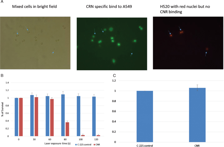
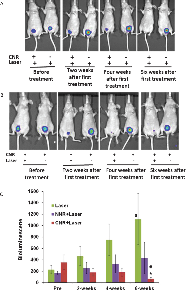
Gold nanorods linked to an anti-EGFR antibody (Conjugated gold NanoRods - CNR) were instilled into the bladder cavity of an orthotopic murine xenograft model with T24 bladder cancer cells expressing luciferase. NIR light was externally administered via an 808 nm diode laser. This treatment was repeated weekly for 4 weeks. The anti-cancer effect was monitored by an imaging system in a non-invasive manner, which was the primary outcome of our study.
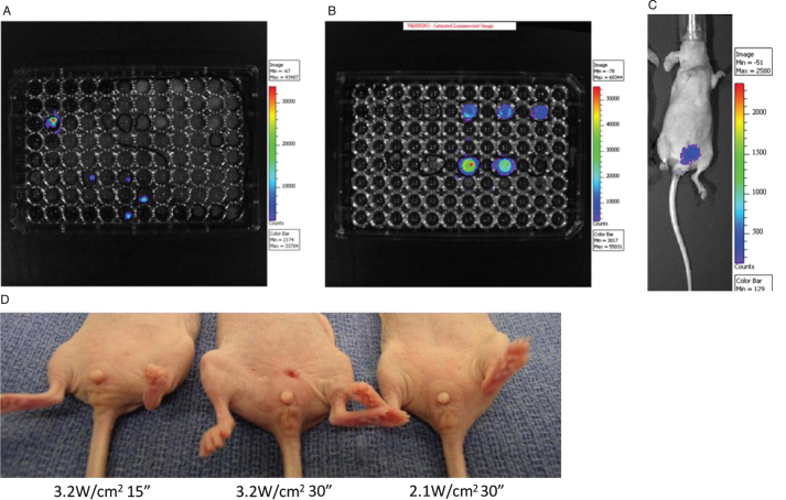
The optimal approach for an individual treatment was 2.1 W/cm laser power for 30 seconds. Using this model, NIR light combined with CNR demonstrated a statistically significant reduction in tumor-associated bioluminescent activity ( = 16) compared to mice treated with laser alone ( = 14) at the end of the study ( = 0.035). Furthermore, the CNR+NIR light treatment significantly abrogated bioluminescence signals over a 6-week observation period, compared to pre-treatment levels ( = 0.045).
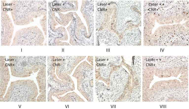
Photothermal tumor ablation with EGFR-directed gold nanorods and NIR light proved effective and well tolerated in a murine model of urinary bladder cancer.
经近红外(NIR)光处理的金纳米颗粒可被优先加热,从而实现对靶向细胞的热消融。将新型膀胱内纳米颗粒导向疗法与通过光纤膀胱镜进行的激光照射相结合,代表了一种针对浅表性膀胱癌患者的潜在消融治疗方法。
在原位膀胱癌模型中,研究用近红外光照射表皮生长因子受体(EGFR)导向的金纳米棒的热消融效果。
将与抗EGFR抗体连接的金纳米棒(共轭金纳米棒 - CNR)注入表达荧光素酶的T24膀胱癌细胞原位小鼠异种移植模型的膀胱腔内。通过808纳米二极管激光从外部施加近红外光。每周重复此治疗,持续4周。通过成像系统以非侵入性方式监测抗癌效果,这是我们研究的主要结果。
个体治疗的最佳方法是2.1瓦/平方厘米的激光功率照射30秒。使用该模型,与仅接受激光治疗的小鼠(n = 14)相比,在研究结束时,近红外光联合CNR显示肿瘤相关生物发光活性有统计学显著降低(n = 16)(P = 0.035)。此外,与治疗前水平相比,在6周观察期内,CNR +近红外光治疗显著消除了生物发光信号(P = 0.045)。
在小鼠膀胱癌模型中,用EGFR导向的金纳米棒和近红外光进行光热肿瘤消融证明是有效的且耐受性良好。