Akiki Teddy J, Averill Christopher L, Wrocklage Kristen M, Schweinsburg Brian, Scott J Cobb, Martini Brenda, Averill Lynnette A, Southwick Steven M, Krystal John H, Abdallah Chadi G
National Center for PTSD - Clinical Neurosciences Division, US Department of Veterans Affairs, West Haven, Connecticut.
Department of Psychiatry, Yale University School of Medicine, New Haven, Connecticut.
Chronic Stress (Thousand Oaks). 2017 Feb;1. doi: 10.1177/2470547017724069. Epub 2017 Aug 8.
The hippocampus and amygdala have been repeatedly implicated in the psychopathology of posttraumatic stress disorder (PTSD). While numerous structural neuroimaging studies examined these two structures in PTSD, these analyses have largely been limited to volumetric measures. Recent advances in vertex-based neuroimaging methods have made it possible to identify specific locations of subtle morphometric changes within a structure of interest.
In this cross-sectional study, we used high-resolution magnetic resonance imaging to examine the relationship between PTSD symptomatology, as measured using the Clinician Administered PTSD Scale for the DSM-IV (CAPS), and structural shape of the hippocampus and amygdala using vertex-wise shape analyses in a group of combat-exposed US Veterans (N = 69).
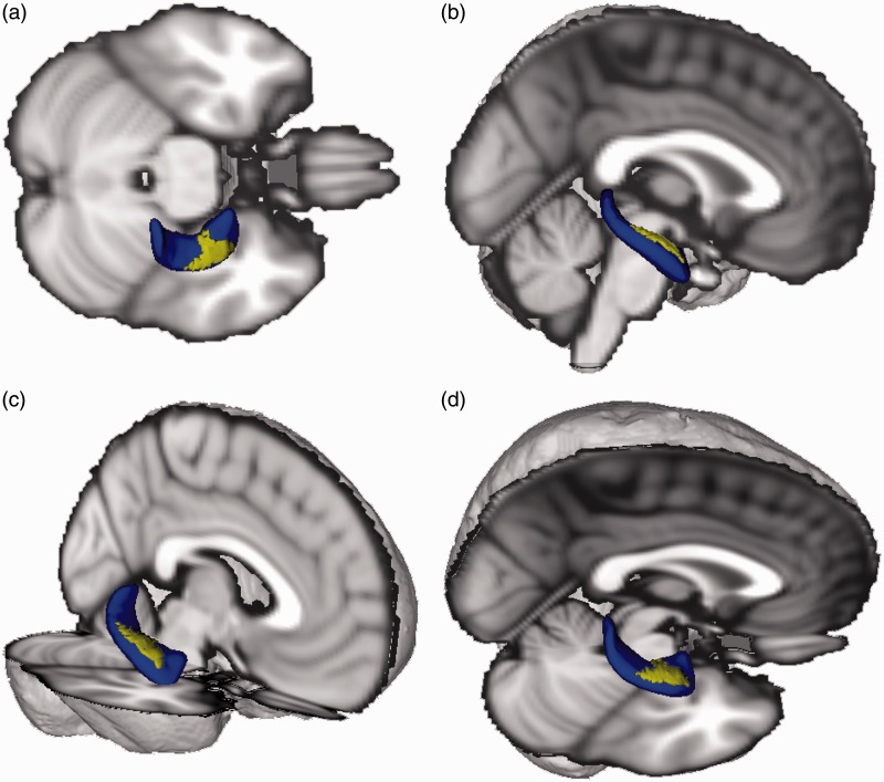
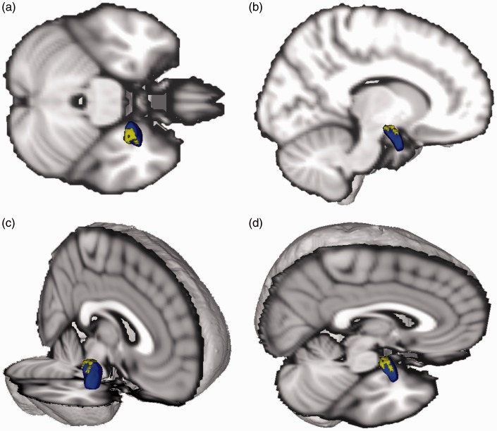
Following correction for multiple comparisons and controlling for age and cranial volume, we found that participants with more severe PTSD symptoms showed an indentation in the anterior half of the right hippocampus and an indentation in the dorsal region of the right amygdala (corresponding to the centromedial amygdala). Post hoc analysis using stepwise regression suggest that among PTSD symptom clusters, arousal symptoms explain most of the variance in the hippocampal abnormality, whereas re-experiencing symptoms explain most of the variance in the amygdala abnormality.
The results provide evidence of localized abnormalities in the anterior hippocampus and centromedial amygdala in combat-exposed US Veterans suffering from PTSD symptoms. This novel finding provides a more fine-grained analysis of structural abnormalities in PTSD and may be informative for understanding the neurobiology of the disorder.
海马体和杏仁核反复被认为与创伤后应激障碍(PTSD)的精神病理学有关。虽然众多结构神经影像学研究在PTSD中对这两个结构进行了检查,但这些分析大多局限于体积测量。基于顶点的神经影像学方法的最新进展使得识别感兴趣结构内细微形态计量变化的特定位置成为可能。
在这项横断面研究中,我们使用高分辨率磁共振成像来检查创伤后应激障碍症状(使用针对《精神疾病诊断与统计手册》第四版的临床医生管理的创伤后应激障碍量表(CAPS)进行测量)与一组有战斗经历的美国退伍军人(N = 69)中海马体和杏仁核的结构形状之间的关系,采用顶点-wise形状分析。
在进行多重比较校正并控制年龄和颅容积后,我们发现创伤后应激障碍症状更严重的参与者右侧海马体前半部分有凹陷,右侧杏仁核背侧区域(对应于中央内侧杏仁核)有凹陷。使用逐步回归的事后分析表明,在创伤后应激障碍症状群中,唤醒症状解释了海马体异常中大部分的变异,而重新体验症状解释了杏仁核异常中大部分的变异。
结果为患有创伤后应激障碍症状的有战斗经历的美国退伍军人的前海马体和中央内侧杏仁核存在局部异常提供了证据。这一新发现为创伤后应激障碍的结构异常提供了更精细的分析,可能有助于理解该疾病的神经生物学机制。