Department of Psychiatry Affiliated Nanjing Brain Hospital of Nanjing Medical University Nanjing China.
Research Center of Learning Science Southeast University Nanjing China.
Brain Behav. 2017 Jun 27;7(8):e00754. doi: 10.1002/brb3.754. eCollection 2017 Aug.
Anxious depression is one of the common subtypes of major depressive disorder (MDD). Clinically, patients with anxious depression exhibit more severe depressive symptoms than patients with nonanxious depression. The aim of the present study was to explore the common and differing cortical and subcortical structural changes between patients with anxious and nonanxious depression.
Patients were placed into one of three groups: the anxious depression group (MDD patients with high levels of anxiety symptoms, = 23), the nonanxious depression group ( = 22), and healthy controls ( = 43) that were matched for age, sex, and education level. All participants underwent T1-weighted MRI. The Freesurfer, which uses a set of automated sequences to analyze the abnormal changes of cortical thickness, cortical and subcortical structures, was used to process the T1 images.
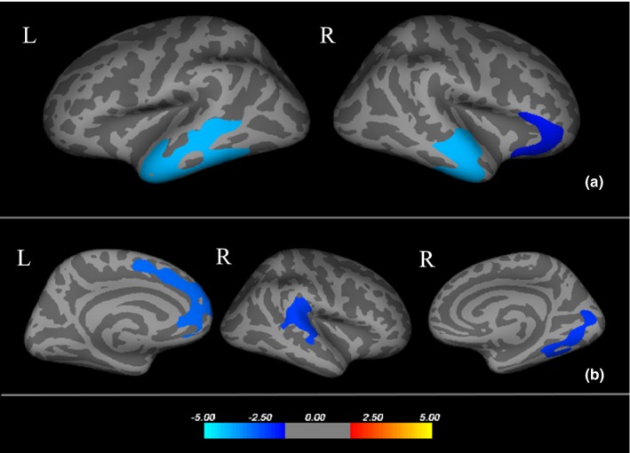
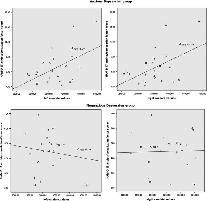
Compared to controls, MDD patients showed thinner cortical thickness in the left inferior temporal, the right superior temporal, and the right parsorbitalis, and a smaller volume of the left hippocampus. Compared to nonanxious depression, anxious depressive patients showed a cortical thinning of the left superior frontal and right superior temporal, as well as the right lingual, and significantly increased subcortical volume of the bilateral caudate nuclei. Correlation analysis showed that the volumes of the bilateral caudate nuclei were directly proportional to the anxiety/somatization factor score.
These findings suggest that smaller hippocampal volume and atrophic prefrontal and temporal cortices might be a common pattern of cortical and subcortical alterations in patients with depression and/or anxiety. However, the change in the caudate nucleus volume may be indicative of anxious depression and may potentially be used to distinguish anxious from nonanxious depression.
焦虑性抑郁症是重性抑郁障碍(MDD)的常见亚型之一。临床上,焦虑性抑郁症患者的抑郁症状比非焦虑性抑郁症患者更为严重。本研究旨在探讨焦虑性和非焦虑性抑郁症患者皮质和皮质下结构的常见和不同变化。
将患者分为三组:焦虑性抑郁症组(焦虑症状水平较高的 MDD 患者,n=23)、非焦虑性抑郁症组(n=22)和健康对照组(n=43),这些组在年龄、性别和教育水平上相匹配。所有参与者均接受 T1 加权 MRI 检查。使用 Freesurfer 分析皮质厚度、皮质和皮质下结构的异常变化,这是一套自动序列,用于处理 T1 图像。
与对照组相比,MDD 患者左颞下回、右颞上回和右眶额皮质的皮质厚度变薄,左海马体积减小。与非焦虑性抑郁症相比,焦虑性抑郁症患者左额上回和右颞上回皮质变薄,以及右舌回皮质变薄,双侧尾状核体积明显增大。相关性分析表明,双侧尾状核体积与焦虑/躯体化因子评分呈正相关。
这些发现表明,较小的海马体积和萎缩的额颞皮质可能是抑郁症和/或焦虑症患者皮质和皮质下改变的共同模式。然而,尾状核体积的变化可能提示焦虑性抑郁症,并且可能潜在地用于区分焦虑性和非焦虑性抑郁症。