Division of Molecular Toxicology, Karolinska Institutet, Stockholm, Sweden.
Division of Biochemical Toxicology, Institute of Environmental Medicine, Karolinska Institutet, Stockholm, Sweden.
Sci Rep. 2017 Aug 24;7(1):9284. doi: 10.1038/s41598-017-09430-8.
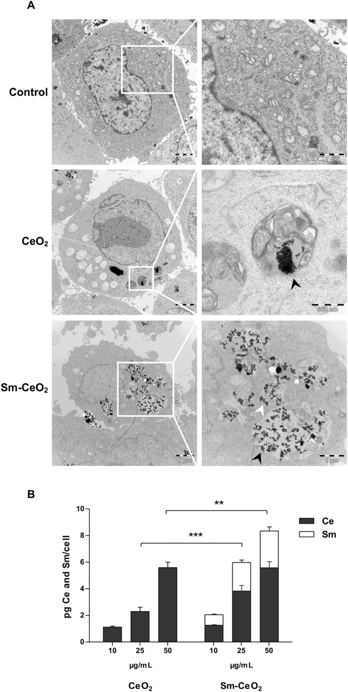
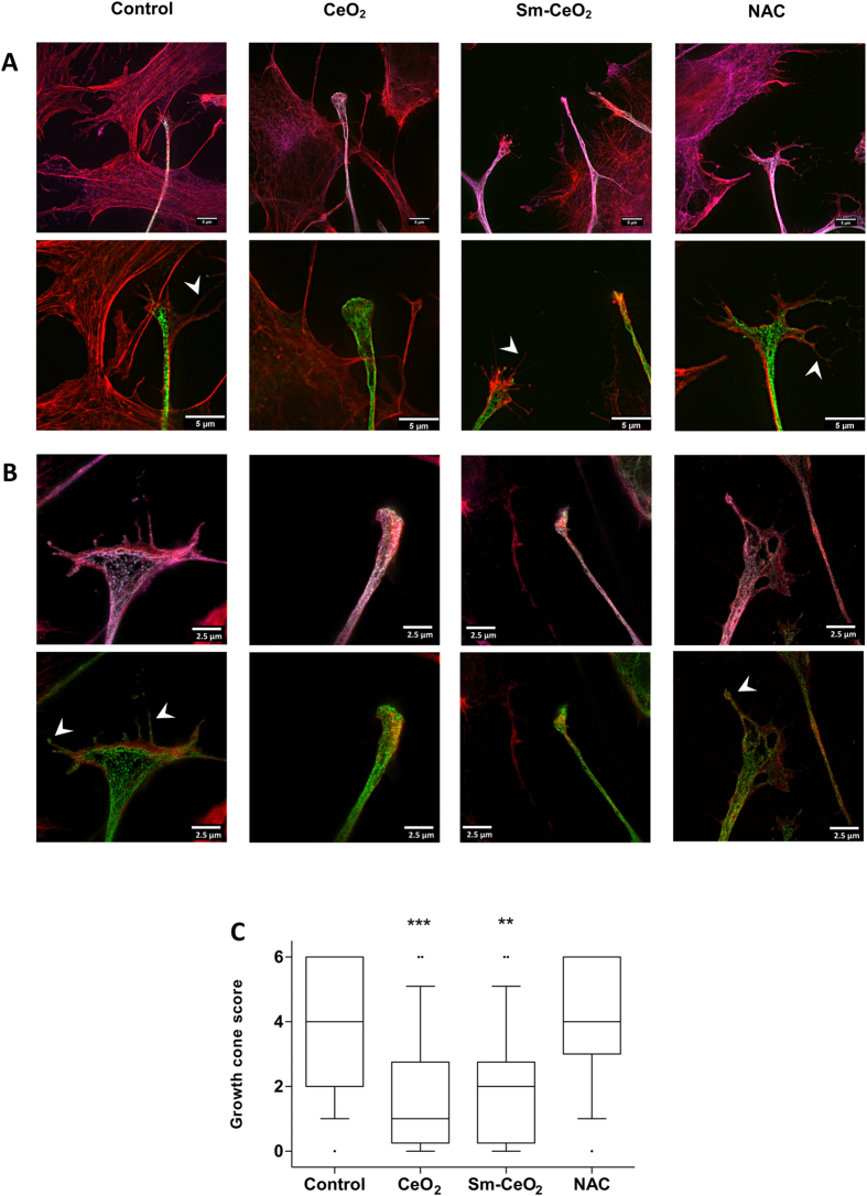
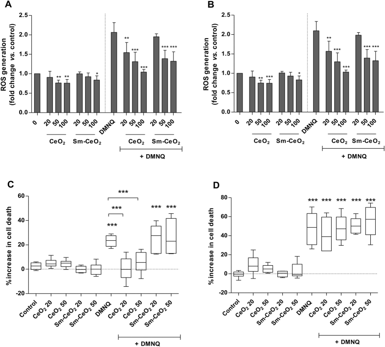
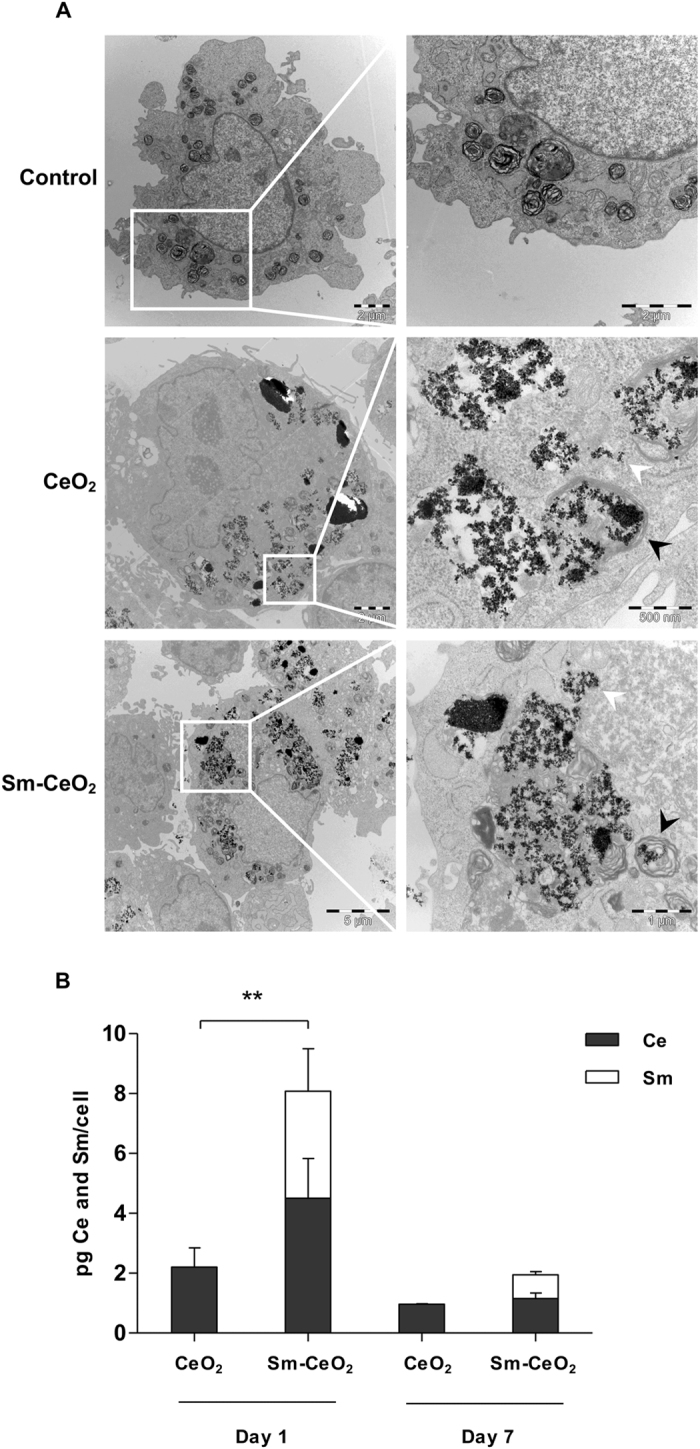
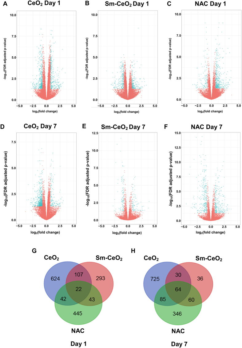
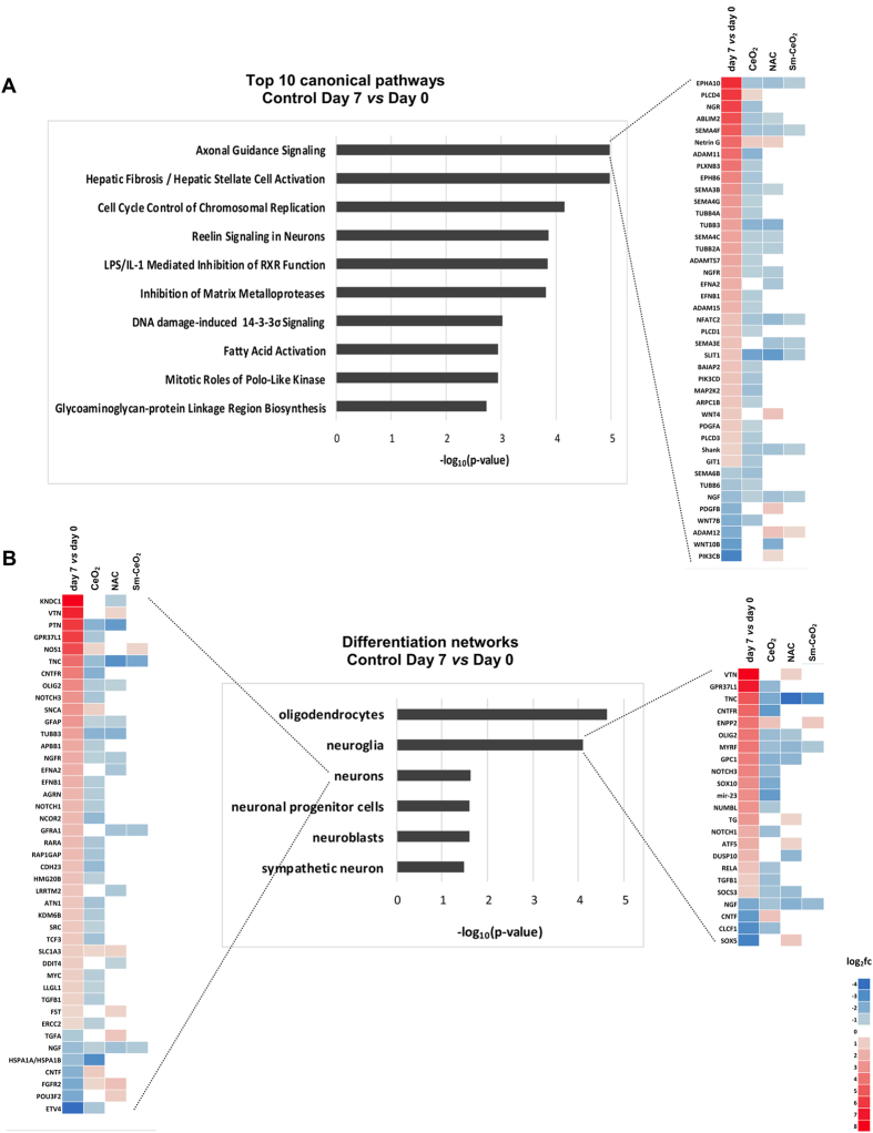
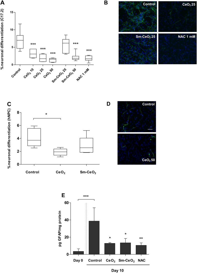
Cerium oxide nanoparticles (nanoceria) display antioxidant properties and have shown cytoprotective effects both in vitro and in vivo. Here, we explored the effects of nanoceria on neural progenitor cells using the C17.2 murine cell line as a model. First, we assessed the effects of nanoceria versus samarium (Sm) doped nanoceria on cell viability in the presence of the prooxidant, DMNQ. Both particles were taken up by cells and nanoceria, but not Sm-doped nanoceria, elicited a temporary cytoprotective effect upon exposure to DMNQ. Next, we employed RNA sequencing to explore the transcriptional responses induced by nanoceria or Sm-doped nanoceria during neuronal differentiation. Detailed computational analyses showed that nanoceria altered pathways and networks relevant for neuronal development, leading us to hypothesize that nanoceria inhibits neuronal differentiation, and that nanoceria and Sm-doped nanoceria both interfere with cytoskeletal organization. We confirmed that nanoceria reduced neuron specific β3-tubulin expression, a marker of neuronal differentiation, and GFAP, a neuroglial marker. Furthermore, using super-resolution microscopy approaches, we could show that both particles interfered with cytoskeletal organization and altered the structure of neural growth cones. Taken together, these results reveal that nanoceria may impact on neuronal differentiation, suggesting that nanoceria could pose a developmental neurotoxicity hazard.
氧化铈纳米颗粒(纳米氧化铈)具有抗氧化特性,并已在体外和体内显示出细胞保护作用。在这里,我们使用 C17.2 小鼠细胞系作为模型,探索了纳米氧化铈对神经祖细胞的影响。首先,我们评估了纳米氧化铈与掺钐(Sm)的纳米氧化铈对存在促氧化剂 DMNQ 时细胞活力的影响。两种颗粒都被细胞摄取,而只有纳米氧化铈而不是掺 Sm 的纳米氧化铈在暴露于 DMNQ 时产生暂时的细胞保护作用。接下来,我们使用 RNA 测序来探索纳米氧化铈或掺 Sm 的纳米氧化铈在神经元分化过程中诱导的转录反应。详细的计算分析表明,纳米氧化铈改变了与神经元发育相关的途径和网络,这使我们假设纳米氧化铈抑制神经元分化,并且纳米氧化铈和掺 Sm 的纳米氧化铈都干扰细胞骨架组织。我们证实纳米氧化铈降低了神经元特异性β3-微管蛋白表达,这是神经元分化的标志物,以及神经胶质标志物 GFAP。此外,使用超分辨率显微镜方法,我们可以表明这两种颗粒都干扰了细胞骨架组织并改变了神经生长锥的结构。总之,这些结果表明纳米氧化铈可能会影响神经元分化,表明纳米氧化铈可能构成发育神经毒性危害。