Wang Tao, Sun Dong, Liu Yumin, Mei Bin, Li Huagang, Zhang Shengming, Zhang Junjian
Department of Neurology, Zhongnan Hospital, Wuhan University, Wuhan, China.
Front Neurol. 2017 Aug 9;8:403. doi: 10.3389/fneur.2017.00403. eCollection 2017.
Asymptomatic carotid artery stenosis can lead to not only stroke but also cognition impairment. Although it has been proven that carotid artery stenting (CAS) can reduce the risk of future strokes, the effect of CAS on cognition is conflicting. In recent years, pulsed arterial spin labeling (pASL) MRI and resting-state functional MRI (R-fMRI) have been employed in cognitive impairment studies. For the present study, cognition is evaluated in severe asymptomatic carotid artery stenosis patients undergoing CAS, and the mechanisms underlying the cognitive change are explored by pASL MRI and R-fMRI.
We prospectively enrolled 24 asymptomatic, severe (≥70%), unilateral internal carotid artery stenosis patients, who were expecting the intervention of CAS. Cognition assessment (including the Montreal Cognitive Assessment Beijing Version, the Minimum Mental State Examination, the Digit Symbol Test, the Rey Auditory Verbal Learning Test, and the Verbal Memory Test) and an integrated MRI program (pASL MRI, and R-fMRI) were administered 7 days before and 3 months after CAS.
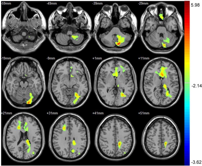
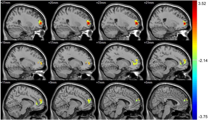
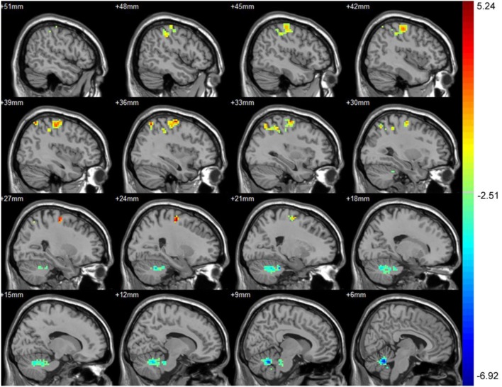
16 subjects completed the follow-up study. After stenting, significant improvement in the scores of the MMSE, the Verbal Memory test, and the delayed recall was found. No significant difference was found in the scores of the Montreal Cognitive Assessment Beijing Version, the Digit Symbol Test, and the immediate recall. After CAS treatment, asymptomatic carotid artery stenosis patients showed increased perfusion in the left frontal gyrus, increased amplitude of low-frequency fluctuation (ALFF) in the right precentral gyrus, and increased connectivity to the posterior cingulate cortex (PCC) in the right supra frontal gyrus. However, no significant correlations were found between these imaging changes and cognition assessments.
Successful CAS can partly improve cognition in asymptomatic carotid artery stenosis patients. The cognition improvement may be partly attributed to the increased perfusion in the left frontal gyrus, increased ALFF in the right precentral gyrus, and increased connectivity to the PCC in the right supra frontal gyrus.
无症状性颈动脉狭窄不仅可导致卒中,还可引起认知功能障碍。尽管已证实颈动脉支架置入术(CAS)可降低未来卒中风险,但CAS对认知功能的影响仍存在争议。近年来,脉冲动脉自旋标记(pASL)磁共振成像(MRI)和静息态功能MRI(R-fMRI)已应用于认知功能障碍研究。在本研究中,对接受CAS治疗的重度无症状性颈动脉狭窄患者的认知功能进行评估,并通过pASL MRI和R-fMRI探索认知改变的潜在机制。
我们前瞻性纳入了24例无症状、重度(≥70%)单侧颈内动脉狭窄患者,这些患者预期接受CAS治疗。在CAS治疗前7天和治疗后3个月进行认知评估(包括蒙特利尔认知评估北京版、简易精神状态检查表、数字符号测验、雷氏听觉词语学习测验和言语记忆测验)以及综合MRI检查(pASL MRI和R-fMRI)。
16例受试者完成了随访研究。支架置入术后,简易精神状态检查表、言语记忆测验和延迟回忆得分有显著改善。蒙特利尔认知评估北京版、数字符号测验和即时回忆得分无显著差异。CAS治疗后,无症状性颈动脉狭窄患者左侧额下回灌注增加,右侧中央前回低频振幅(ALFF)增加,右侧额上回与后扣带回皮质(PCC)的连接性增加。然而,这些影像学改变与认知评估之间未发现显著相关性。
成功的CAS可部分改善无症状性颈动脉狭窄患者的认知功能。认知功能的改善可能部分归因于左侧额下回灌注增加、右侧中央前回ALFF增加以及右侧额上回与PCC连接性增加。