Research Institute of Ophthalmology and Visual Sciences, Affiliated Eye Hospital of Nanchang University, Nanchang, Jiangxi 330006, P.R. China.
Department of Pharmacology, School of Pharmaceutical Sciences, Nanchang University, Nanchang, Jiangxi 330006, P.R. China.
Mol Med Rep. 2017 Oct;16(4):4521-4528. doi: 10.3892/mmr.2017.7203. Epub 2017 Aug 10.
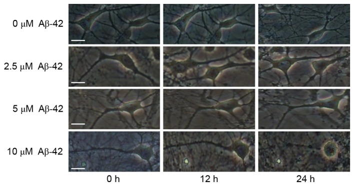
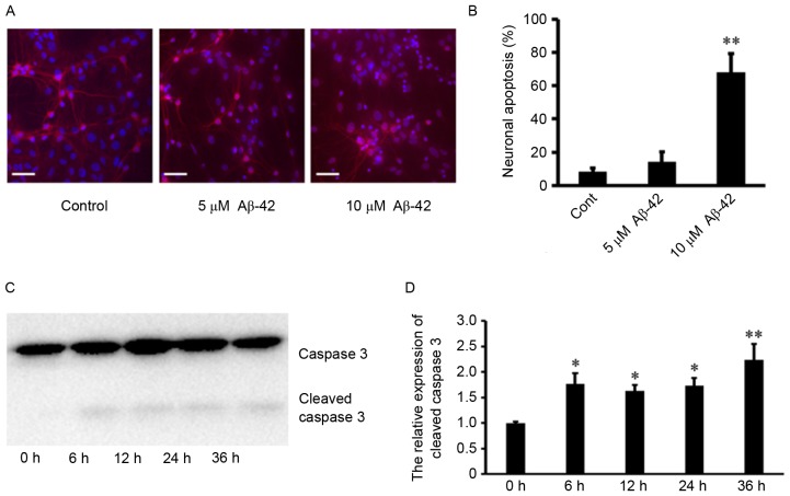
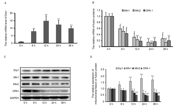
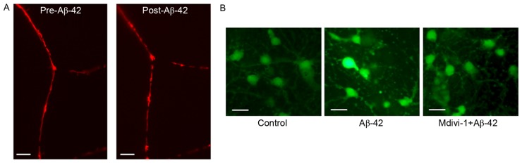
Alzheimer's disease (AD), with a typical pathological hallmark of amyloid‑beta (Aβ)‑containing plaques and neurofibrillary tangles, is one of the most common types of chronic neurodegenerative diseases. Aβ oligomers serve a crucial role in the pathogenesis of AD, and lead to neuronal loss. However, the precise mechanism of Aβ oligomers in AD remains to be elucidated. The present study demonstrated that 10 µM Aβ‑42 activated the caspase signaling pathway, and induced significant apoptosis in primary cultured mouse cerebral cortical neurons. The results of reverse transcription‑quantitative polymerase chain reaction and western blotting demonstrated that Aβ‑42 (10 µM) also significantly upregulated the transcription and expression of the mitochondrial fission protein dynamin‑related protein 1 (Drp1), and downregulated the transcription and expression of mitochondrial fusion proteins, including mitofusin 1/2 (Mfn1/2) and mitochondrial dynamin like GTPase (OPA‑1). Neurons were transfected with pDsRed2‑Mito for mitochondrial imaging, which revealed that 10 µM Aβ‑42 induced mitochondrial fission in cortical neurons. In addition, 2',7'‑dichlorodihydrofluorescein diacetate and tetramethylrhodamine ethyl ester staining indicated that Aβ‑42 increased the reactive oxygen species (ROS) level and reduced mitochondrial membrane potential in neurons. Inhibition of Drp1 activity by Mdivi‑1 efficiently prevented Aβ‑42‑induced ROS production and disruption of mitochondrial membrane potential. Loss of mitochondrial membrane potential may activate PTEN‑induced putative kinase 1 (Pink1), the prominent sensor for mitochondrial damage, and trigger the process of mitophagy to remove the damaged mitochondria. In the present study, western blotting revealed that the levels of autophagy marker microtubule‑associated proteins 1A/1B light chain 3B (LC3B) and Pink1 were upregulated after Aβ‑42 stimulation. In conclusion, these data indicated that Aβ‑42 induces neuronal apoptosis by targeting mitochondria, including promotion of mitochondrial fission, disruption of mitochondrial membrane potential, increasing intracellular ROS level and activation of the process of mitophagy. Therefore, mitochondria may represent a potential therapeutic target for AD in the future.
阿尔茨海默病(AD)的典型病理学特征是含有淀粉样β(Aβ)的斑块和神经原纤维缠结,是最常见的慢性神经退行性疾病之一。Aβ寡聚体在 AD 的发病机制中起着至关重要的作用,并导致神经元丢失。然而,Aβ寡聚体在 AD 中的确切机制仍有待阐明。本研究表明,10 μM Aβ-42 激活了半胱天冬酶信号通路,并诱导原代培养的小鼠大脑皮质神经元发生显著凋亡。逆转录-定量聚合酶链反应和 Western blot 结果表明,Aβ-42(10 μM)还显著上调了线粒体分裂蛋白动力相关蛋白 1(Drp1)的转录和表达,并下调了线粒体融合蛋白,包括线粒体融合蛋白 1/2(Mfn1/2)和线粒体动力相关 GTP 酶(OPA-1)的转录和表达。用 pDsRed2-Mito 转染神经元进行线粒体成像,结果显示 10 μM Aβ-42 诱导皮质神经元发生线粒体分裂。此外,2',7'-二氯二氢荧光素二乙酸酯和四甲基罗丹明乙酯染色表明,Aβ-42 增加了神经元中的活性氧(ROS)水平并降低了线粒体膜电位。用 Mdivi-1 抑制 Drp1 活性可有效防止 Aβ-42 诱导的 ROS 产生和线粒体膜电位破坏。线粒体膜电位的丧失可能会激活 PTEN 诱导的假定激酶 1(Pink1),这是线粒体损伤的主要传感器,并触发自噬过程以清除受损的线粒体。在本研究中,Western blot 显示 Aβ-42 刺激后自噬标志物微管相关蛋白 1A/1B 轻链 3B(LC3B)和 Pink1 的水平上调。总之,这些数据表明,Aβ-42 通过靶向线粒体诱导神经元凋亡,包括促进线粒体分裂、破坏线粒体膜电位、增加细胞内 ROS 水平和激活自噬过程。因此,线粒体可能是未来 AD 的潜在治疗靶点。