Melbourne Neuropsychiatry Centre, Department of Psychiatry, The University of Melbourne and Melbourne Health, Carlton South, VIC, Australia.
Department of Psychiatry, The University of Melbourne, Parkville, VIC Australia.
Transl Psychiatry. 2017 Aug 29;7(8):e1225. doi: 10.1038/tp.2017.193.
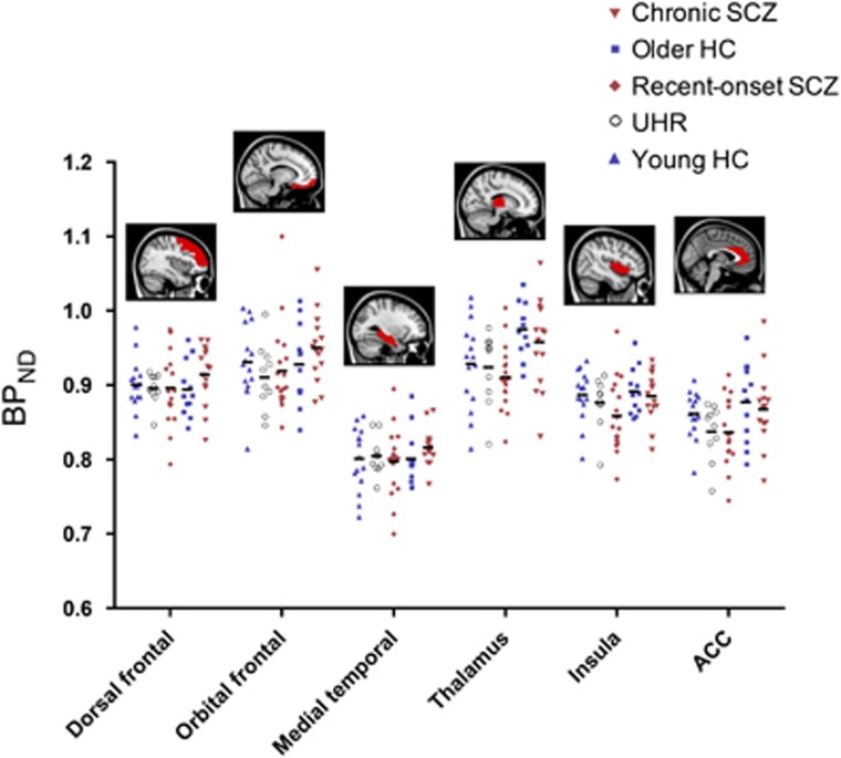
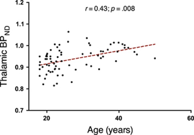
We examined putative microglial activation as a function of illness course in schizophrenia. Microglial activity was quantified using C-(1-[2-chrorophynyl]-N-methyl-N-[1-methylpropyl]-3 isoquinoline carboxamide (C-(R)-PK11195) positron emission tomography (PET) in: (i) 10 individuals at ultra-high risk (UHR) of psychosis; (ii) 18 patients recently diagnosed with schizophrenia; (iii) 15 patients chronically ill with schizophrenia; and, (iv) 27 age-matched healthy controls. Regional-binding potential (BP) was calculated using the simplified reference-tissue model with four alternative reference inputs. The UHR, recent-onset and chronic patient groups were compared to age-matched healthy control groups to examine between-group BP differences in 6 regions: dorsal frontal, orbital frontal, anterior cingulate, medial temporal, thalamus and insula. Correlation analysis tested for BP associations with gray matter volume, peripheral cytokines and clinical variables. The null hypothesis of equality in BP between patients (UHR, recent-onset and chronic) and respective healthy control groups (younger and older) was not rejected for any group comparison or region. Across all subjects, BP was positively correlated to age in the thalamus (r=0.43, P=0.008, false discovery rate). No correlations with regional gray matter, peripheral cytokine levels or clinical symptoms were detected. We therefore found no evidence of microglial activation in groups of individuals at high risk, recently diagnosed or chronically ill with schizophrenia. While the possibility of C-(R)-PK11195-binding differences in certain patient subgroups remains, the patient cohorts in our study, who also displayed normal peripheral cytokine profiles, do not substantiate the assumption of microglial activation in schizophrenia as a regular and defining feature, as measured by C-(R)-PK11195 BP.
我们研究了精神分裂症患者疾病进程中潜在的小胶质细胞激活情况。使用C-(1-[2-氯苯基]-N-甲基-N-[1-甲基丙基]-3 异喹啉羧酰胺(C-(R)-PK11195)正电子发射断层扫描(PET),在以下人群中定量测量小胶质细胞活性:(i)10 名处于精神病超高风险(UHR)的个体;(ii)18 名近期诊断为精神分裂症的患者;(iii)15 名患有慢性精神分裂症的患者;以及(iv)27 名年龄匹配的健康对照者。使用简化参考组织模型和四个替代参考输入,计算了各区域的结合势(BP)。将 UHR、近期发病和慢性患者组与年龄匹配的健康对照组进行比较,以检查 6 个区域(背侧额叶、眶额部、前扣带、内侧颞叶、丘脑和脑岛)的组间 BP 差异。相关性分析检验了 BP 与灰质体积、外周细胞因子和临床变量之间的关联。在任何组间比较或区域中,均未拒绝患者(UHR、近期发病和慢性)和相应的健康对照组(年轻和年长)之间的 BP 相等的零假设。在所有受试者中,BP 与丘脑的年龄呈正相关(r=0.43,P=0.008,假发现率)。未检测到与区域性灰质、外周细胞因子水平或临床症状的相关性。因此,我们在处于精神病超高风险、近期发病或慢性精神分裂症的个体组中未发现小胶质细胞激活的证据。虽然在某些患者亚组中存在 C-(R)-PK11195 结合差异的可能性,但在我们的研究中,患者队列也显示出正常的外周细胞因子谱,这不能证明精神分裂症中小胶质细胞激活是一种常规和确定的特征,如 C-(R)-PK11195 BP 所测量的那样。