Zhang Y, Catts V S, Sheedy D, McCrossin T, Kril J J, Shannon Weickert C
Schizophrenia Research Institute, Randwick, NSW, Australia.
Schizophrenia Research Laboratory, Neuroscience Research Australia, Randwick, NSW, Australia.
Transl Psychiatry. 2016 Dec 13;6(12):e982. doi: 10.1038/tp.2016.238.
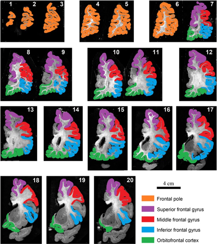
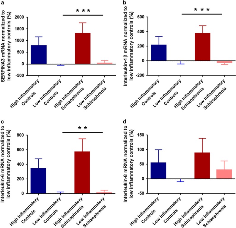
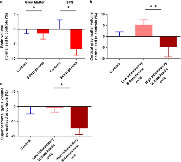
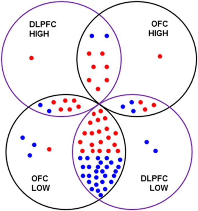
Cortical grey matter volume deficits and neuro-inflammation exist in patients with schizophrenia, although it is not clear whether elevated cytokines contribute to the cortical volume reduction. We quantified cortical and regional brain volumes in fixed postmortem brains from people with schizophrenia and matched controls using stereology. Interleukin (IL)-6, IL-1β, IL-8 and SERPINA3 messenger RNAs (mRNAs) were quantified in the contralateral fresh frozen orbitofrontal cortex. We found a small, but significant reduction in cortical grey matter (1.3%; F(1,85)=4.478, P=0.037) and superior frontal gyrus (6.5%; F(1,80)=5.700, P=0.019) volumes in individuals with schizophrenia compared with controls. Significantly reduced cortical grey matter (9.2%; F(1,24)=8.272, P=0.008) and superior frontal gyrus (13.9%; F(1,20)=5.374, P=0.031) volumes were found in cases with schizophrenia and 'high inflammation' status relative to schizophrenia cases with 'low inflammation' status in the prefrontal cortex. The expression of inflammatory mRNAs in the orbitofrontal cortex was significantly correlated with those in dorsolateral prefrontal cortex (all r>0.417, all P<0.022), except for IL-8. Moreover, average daily and lifetime antipsychotic intake negatively correlated with cortical grey matter and superior frontal gyrus volumes (all r<-0.362, all P<0.05). The results suggest that the reduction in cortical grey matter volume in people with schizophrenia is exaggerated in those who have high expression of inflammatory cytokines. Further, antipsychotic medication intake does not appear to ameliorate the reduction in brain volume.
精神分裂症患者存在皮质灰质体积减少和神经炎症,尽管尚不清楚细胞因子升高是否导致皮质体积减小。我们使用体视学方法对精神分裂症患者和匹配对照者的固定尸检大脑中的皮质和区域脑体积进行了量化。在对侧新鲜冷冻的眶额皮质中对白细胞介素(IL)-6、IL-1β、IL-8和丝氨酸蛋白酶抑制剂A3信使核糖核酸(mRNA)进行了量化。我们发现,与对照组相比,精神分裂症患者的皮质灰质体积(1.3%;F(1,85)=4.478,P=0.037)和额上回体积(6.5%;F(1,80)=5.700,P=0.019)有小幅但显著的减少。与前额叶皮质中“低炎症”状态的精神分裂症病例相比,“高炎症”状态的精神分裂症病例的皮质灰质体积(9.2%;F(1,24)=8.272,P=0.008)和额上回体积(13.9%;F(1,20)=5.374,P=0.031)显著减少。眶额皮质中炎症mRNA的表达与背外侧前额叶皮质中的表达显著相关(所有r>0.417,所有P<0.022),IL-8除外。此外,平均每日和终生抗精神病药物摄入量与皮质灰质和额上回体积呈负相关(所有r<-0.362,所有P<0.05)。结果表明,炎症细胞因子高表达的精神分裂症患者皮质灰质体积的减少更为明显。此外,抗精神病药物治疗似乎并未改善脑体积的减少。