Falkenius Johan, Johansson Hemming, Tuominen Rainer, Frostvik Stolt Marianne, Hansson Johan, Egyhazi Brage Suzanne
Department of Oncology-Pathology, Karolinska Institutet, Cancer Center Karolinska, Karolinska University Hospital, 171 76, Solna, Stockholm, Sweden.
BMC Cancer. 2017 Aug 29;17(1):584. doi: 10.1186/s12885-017-3577-x.
The variable prognosis in stage III cutaneous melanoma is partially due to unknown prognostic factors. Improved prognostic tools are required to define patients with an increased risk of developing metastatic disease who might benefit from adjuvant therapies. The aim was to examine if cellular immune markers in association with tumor proliferation rate and BRAF mutation status have an impact on prognosis in stage III melanoma.
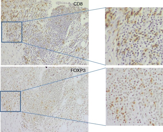
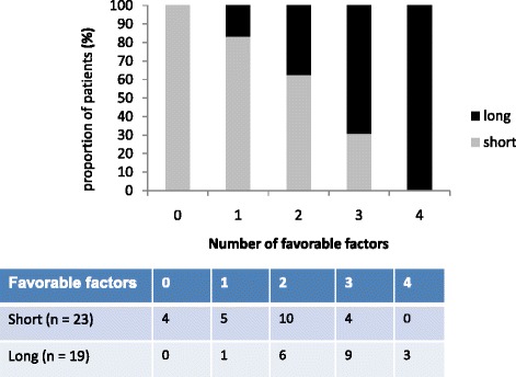
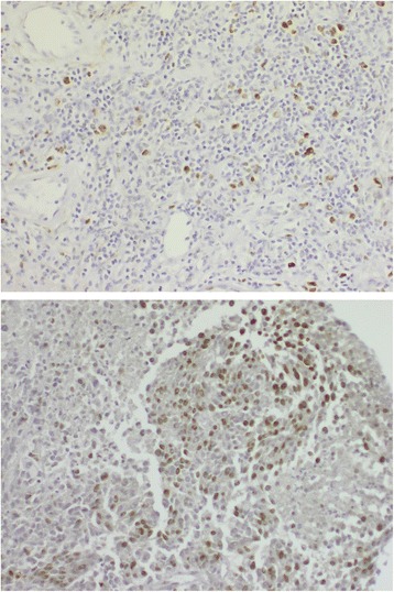
We have used two sets of case series with stage III disease: 23 patients with short survival (≤ 13 months) and 19 patients with long survival (≥ 60 months). Lymph node metastases were analyzed for Ki67, CD8 and FOXP3 protein expression using immunohistochemistry. BRAF mutation status was analyzed in a previous study on the same samples.
Low tumor proliferation rate was significantly associated with a better prognosis (p = 0.013). Presence of FOXP3+ T cells was not correlated to adverse clinical outcome. A highly significant trend for a longer survival was found in the presence of an increasing number of markers; CD8+ and FOXP3+ T cells, low tumor proliferation and BRAF wildtype status (p = 0.003). Presence of at least three of these four markers was found to be an independent favorable prognostic factor (OR 19.4, 95% CI 1.9-197, p = 0.012), when adjusting for ulceration and number of lymph node metastases. Proliferation alone remained significant in multivariate analyses (OR 26.1, 95% CI 2.0-344, p = 0.013) but with a wider confidence interval. This panel still remained independent when also adjusting for a previously identified prognostic glycolytic-pigment panel.
We have demonstrated that presence of immune cells in association with tumor proliferation and BRAF mutation status may further contribute to identify stage III melanoma patients with high risk of relapse.
III期皮肤黑色素瘤预后各异,部分原因是存在未知的预后因素。需要改进预后工具,以确定那些发生转移性疾病风险增加、可能从辅助治疗中获益的患者。本研究旨在探讨细胞免疫标志物与肿瘤增殖率及BRAF突变状态相结合是否会对III期黑色素瘤的预后产生影响。
我们使用了两组III期病例系列:23例生存期短(≤13个月)的患者和19例生存期长(≥60个月)的患者。采用免疫组化法分析淋巴结转移灶中Ki67、CD8和FOXP3蛋白的表达情况。BRAF突变状态在之前对相同样本的研究中已进行分析。
低肿瘤增殖率与较好的预后显著相关(p = 0.013)。FOXP3+ T细胞的存在与不良临床结局无关。随着CD8+和FOXP3+ T细胞数量增加、肿瘤增殖率降低以及BRAF野生型状态的出现,生存期延长的趋势非常显著(p = 0.003)。在调整溃疡和淋巴结转移数量后,发现这四种标志物中至少存在三种是独立的有利预后因素(OR 19.4,95% CI 1.9 - 197,p = 0.012)。在多变量分析中,仅增殖情况仍具有显著性(OR 26.1,95% CI 2.0 - 344,p = 0.013),但置信区间更宽。当同时调整先前确定的预后糖酵解 - 色素指标时,该指标组合仍保持独立性。
我们已经证明,免疫细胞的存在与肿瘤增殖及BRAF突变状态相结合,可能有助于进一步识别具有高复发风险的III期黑色素瘤患者。