文献检索文档翻译深度研究
Suppr Zotero 插件Zotero 插件
邀请有礼套餐&价格历史记录

新学期,新优惠

限时优惠:9月1日-9月22日

30天高级会员仅需29元

1天体验卡首发特惠仅需5.99元

了解详情
不再提醒
插件&应用
Suppr Zotero 插件Zotero 插件浏览器插件Mac 客户端Windows 客户端微信小程序
高级版
套餐订阅购买积分包
AI 工具
文献检索文档翻译深度研究
关于我们
关于 Suppr公司介绍联系我们用户协议隐私条款
关注我们

Suppr 超能文献

核心技术专利:CN118964589B侵权必究
粤ICP备2023148730 号-1Suppr @ 2025

CTA 点征对脑出血患者血肿扩大的预测价值。

Predictive Value of CTA Spot Sign on Hematoma Expansion in Intracerebral Hemorrhage Patients.

机构信息

Department of Neurology, The First Affiliated Hospital of Chongqing Medical University, 1 Youyi Road, Chongqing 400016, China.

Department of Physiology and Pharmacology, Loma Linda University School of Medicine, 11041 Campus Street, Risley Hall, Room 219, Loma Linda, CA 92354, USA.

出版信息

Biomed Res Int. 2017;2017:4137210. doi: 10.1155/2017/4137210. Epub 2017 Aug 9.


DOI:10.1155/2017/4137210
PMID:28852647
原文链接:https://pmc.ncbi.nlm.nih.gov/articles/PMC5567448/
Abstract

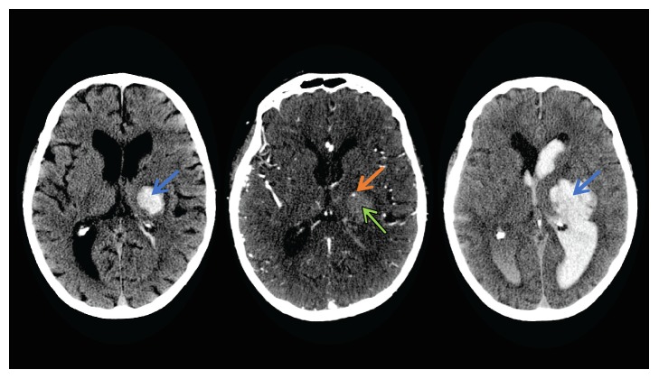
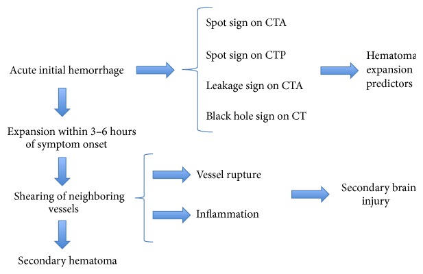
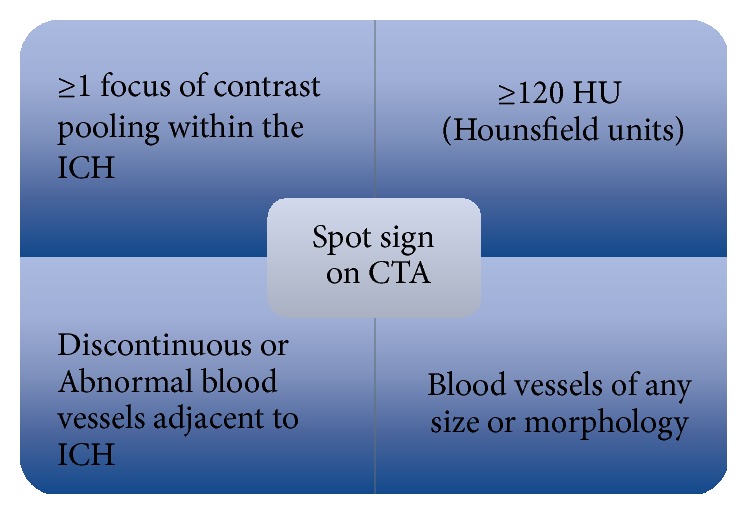
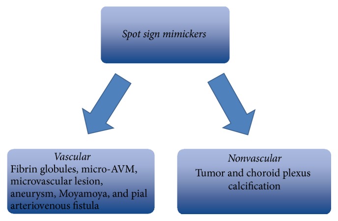
Hematoma expansion (HE) occurs in approximately one-third of patients with intracerebral hemorrhage and leads to high rates of mortality and morbidity. Currently, contrast extravasation within hematoma, termed the spot sign on computed tomography angiography (CTA), has been identified as a strong independent predictor of early hematoma expansion. Past studies indicate that the spot sign is a dynamic entity and is indicative of active hemorrhage. Furthermore, to enhance the spot sign's accuracy of predicting HE, spot parameters observed on CTA or dynamic CTA were used for its quantification. In addition, spot signs detected on multiphase CTA and dynamic CTA are shown to have higher sensitivity and specificity when compared with simple standardized spot sign detection in recent studies. Based on the spot sign, novel methods such as leakage sign and rate of contrast extravasation were explored to redefine HE prediction in combination with clinical characteristics and spot sign on CTA to assist clinical judgment. The spot sign is an accepted independent predictor of active hemorrhage and is used in both secondary intracerebral hemorrhage and the process of surgical assessment for hemorrhagic risk in patients with ischemic stroke. Spot sign predicts patients at high risk for hematoma expansion.

摘要

血肿扩大(HE)约发生于三分之一的脑出血患者中,导致高死亡率和高发病率。目前,血肿内的对比剂外渗,在 CT 血管造影(CTA)上被称为斑点征,已被确定为早期血肿扩大的强独立预测因子。过去的研究表明,斑点征是一个动态实体,提示有活跃性出血。此外,为了提高斑点征预测 HE 的准确性,使用 CTA 或动态 CTA 上观察到的斑点参数对其进行定量。此外,与简单的标准化斑点征检测相比,多期 CTA 和动态 CTA 上检测到的斑点征在最近的研究中显示出更高的敏感性和特异性。基于斑点征,一些新的方法如漏出征和对比剂外渗率被探索出来,以结合临床特征和 CTA 上的斑点征来重新定义 HE 预测,以辅助临床判断。斑点征是活跃性出血的公认独立预测因子,用于继发性脑出血和缺血性脑卒中患者的手术评估中出血风险的过程。斑点征预测血肿扩大风险高的患者。

https://cdn.ncbi.nlm.nih.gov/pmc/blobs/9fab/5567448/bdf658972b66/BMRI2017-4137210.004.jpg
https://cdn.ncbi.nlm.nih.gov/pmc/blobs/9fab/5567448/fabb8844c38b/BMRI2017-4137210.001.jpg
https://cdn.ncbi.nlm.nih.gov/pmc/blobs/9fab/5567448/d42e927ede32/BMRI2017-4137210.002.jpg
https://cdn.ncbi.nlm.nih.gov/pmc/blobs/9fab/5567448/dbb045cc6861/BMRI2017-4137210.003.jpg
https://cdn.ncbi.nlm.nih.gov/pmc/blobs/9fab/5567448/bdf658972b66/BMRI2017-4137210.004.jpg

相似文献

[1]
Predictive Value of CTA Spot Sign on Hematoma Expansion in Intracerebral Hemorrhage Patients.

Biomed Res Int. 2017-8-9

[2]
Rate of Contrast Extravasation on Computed Tomographic Angiography Predicts Hematoma Expansion and Mortality in Primary Intracerebral Hemorrhage.

Stroke. 2015-9

[3]
Predicting Intracerebral Hemorrhage Growth With the Spot Sign: The Effect of Onset-to-Scan Time.

Stroke. 2016-3

[4]
Added value of delayed computed tomography angiography in primary intracranial hemorrhage and hematoma size for predicting spot sign.

Acta Radiol. 2018-4

[5]
Significance of satellite sign and spot sign in predicting hematoma expansion in spontaneous intracerebral hemorrhage.

Clin Neurol Neurosurg. 2017-11

[6]
Prediction of haematoma growth and outcome in patients with intracerebral haemorrhage using the CT-angiography spot sign (PREDICT): a prospective observational study.

Lancet Neurol. 2012-3-8

[7]
Venous phase of computed tomography angiography increases spot sign detection, but intracerebral hemorrhage expansion is greater in spot signs detected in arterial phase.

Stroke. 2014-1-30

[8]
Eliminating vascular interference from the Spot Sign contributes to predicting hematoma expansion in individuals with spontaneous cerebral hemorrhages.

Acta Neurol Belg. 2021-4

[9]
CT Angiography Spot Sign, Hematoma Expansion, and Outcome in Primary Pontine Intracerebral Hemorrhage.

Neurocrit Care. 2016-8

[10]
Prevalence and the predictive performance of the dynamic CT-angiography spot sign in an observational cohort with intracerebral hemorrhage.

Medicine (Baltimore). 2020-11-20

引用本文的文献

[1]
Imaging predictors of hemorrhagic progression of a contusion after traumatic brain injury: a systematic review and meta-analysis.

Sci Rep. 2024-3-12

[2]
Ferroptosis and mitochondrial dysfunction in acute central nervous system injury.

Front Cell Neurosci. 2023-8-9

[3]
Prediction of Hematoma Expansion in Hypertensive Intracerebral Hemorrhage by a Radiomics Nomogram.

Pak J Med Sci. 2023

[4]
A predictive nomogram for intracerebral hematoma expansion based on non-contrast computed tomography and clinical features.

Neuroradiology. 2022-8

[5]
Comparison of Radiomic Models Based on Different Machine Learning Methods for Predicting Intracerebral Hemorrhage Expansion.

Clin Neuroradiol. 2022-3

[6]
Quantitative CT radiomics-based models for prediction of haematoma expansion and poor functional outcome in primary intracerebral haemorrhage.

Eur Radiol. 2021-10

[7]
Is four-dimensional CT angiography as effective as digital subtraction angiography in the detection of the underlying causes of intracerebral haemorrhage: a systematic review.

Neuroradiology. 2020-1-4

[8]
Correlation between Spot Sign and Intracranial Hemorrhage Expansion on Dual-Phase CT Angiography.

Diagnostics (Basel). 2019-12-7

[9]
The risk factors and prognosis of delayed perihematomal edema in patients with spontaneous intracerebral hemorrhage.

CNS Neurosci Ther. 2019-9-22

[10]
Noncontrast computer tomography-based radiomics model for predicting intracerebral hemorrhage expansion: preliminary findings and comparison with conventional radiological model.

Eur Radiol. 2019-8-5

本文引用的文献

[1]
Intracerebral Hemorrhage: Perihemorrhagic Edema and Secondary Hematoma Expansion: From Bench Work to Ongoing Controversies.

Front Neurol. 2016-11-21

[2]
Role of prothrombin complex concentrate (PCC) in Acute Intracerebral Hemorrhage with Positive CTA spot sign: An institutional experience at a regional and state designated stroke center.

Emerg Radiol. 2017-6

[3]
Management of acute stroke in patients on oral anticoagulants.

Curr Opin Neurol. 2017-2

[4]
The critical care management of spontaneous intracranial hemorrhage: a contemporary review.

Crit Care. 2016-9-18

[5]
Negative spot sign in primary intracerebral hemorrhage: potential impact in reducing imaging.

Emerg Radiol. 2017-2

[6]
Association Between Hypodensities Detected by Computed Tomography and Hematoma Expansion in Patients With Intracerebral Hemorrhage.

JAMA Neurol. 2016-8-1

[7]
Black Hole Sign: Novel Imaging Marker That Predicts Hematoma Growth in Patients With Intracerebral Hemorrhage.

Stroke. 2016-7

[8]
Timing of Occurrence Is the Most Important Characteristic of Spot Sign.

Stroke. 2016-5

[9]
Leakage Sign for Primary Intracerebral Hemorrhage: A Novel Predictor of Hematoma Growth.

Stroke. 2016-4

[10]
Predicting Intracerebral Hemorrhage Growth With the Spot Sign: The Effect of Onset-to-Scan Time.

Stroke. 2016-3

文献AI研究员

20分钟写一篇综述,助力文献阅读效率提升50倍

立即体验

用中文搜PubMed

大模型驱动的PubMed中文搜索引擎

马上搜索

推荐工具

医学文档翻译智能文献检索